. 2016 Mar 25;291(13):7156-70.
doi: 10.1074/jbc.M115.686907. Epub 2016 Jan 27.

Post-endocytotic Deubiquitination and Degradation of the Metabotropic γ-Aminobutyric Acid Receptor by the Ubiquitin-specific Protease 14

Affiliations

Post-endocytotic Deubiquitination and Degradation of the Metabotropic γ-Aminobutyric Acid Receptor by the Ubiquitin-specific Protease 14

Nicolas Lahaie et al. J Biol Chem. .

Abstract

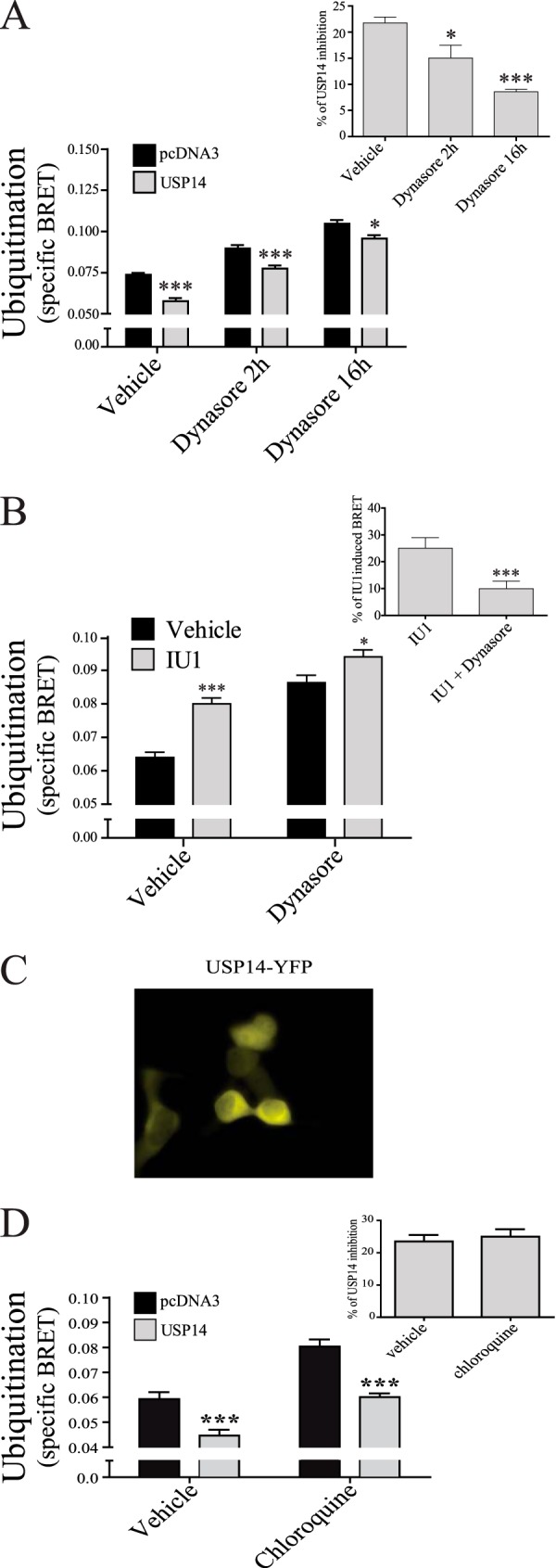
Mechanisms controlling the metabotropic γ-aminobutyric acid receptor (GABAB) cell surface stability are still poorly understood. In contrast with many other G protein-coupled receptors (GPCR), it is not subject to agonist-promoted internalization, but is constitutively internalized and rapidly down-regulated. In search of novel interacting proteins regulating receptor fate, we report that the ubiquitin-specific protease 14 (USP14) interacts with the GABAB(1b)subunit's second intracellular loop. Probing the receptor for ubiquitination using bioluminescence resonance energy transfer (BRET), we detected a constitutive and phorbol 12-myristate 13-acetate (PMA)-induced ubiquitination of the receptor at the cell surface. PMA also increased internalization and accelerated receptor degradation. Overexpression of USP14 decreased ubiquitination while treatment with a small molecule inhibitor of the deubiquitinase (IU1) increased receptor ubiquitination. Treatment with the internalization inhibitor Dynasore blunted both USP14 and IU1 effects on the receptor ubiquitination state, suggesting a post-endocytic site of action. Overexpression of USP14 also led to an accelerated degradation of GABABin a catalytically independent fashion. We thus propose a model whereby cell surface ubiquitination precedes endocytosis, after which USP14 acts as an ubiquitin-binding protein that targets the ubiquitinated receptor to lysosomal degradation and promotes its deubiquitination.

Keywords: G protein-coupled receptor (GPCR); GABA receptor; bioluminescence resonance energy transfer (BRET); biosensor; deubiquitylation (deubiquitination); protein degradation; receptor endocytosis; ubiquitylation (ubiquitination).


Figures

FIGURE 1.
FIGURE 1.
GABAB interacts with USP14 through its GABAB(1) subunit. A, schematic representation of the GABAB(1) second intracellular loop used as bait and the USP14 region recovered in a rat brain cDNA yeast two-hybrid screening. B, HEK293T cells expressing a combination of Myc-GABAB(1b), HA-GABAB(2), Rluc-USP14, and/or Rluc-USP14-C79A were solubilized and immunoprecipitated (IP) using an anti-myc antibody. Total (input, 5% of IP) and IP samples were separated on SDS-PAGE and proteins detected by Western blotting (WB) using the indicated antibodies (anti-myc for GABAB(1b), anti GABAB(2) for GABAB(2), and anti-Rluc for USP14). C, HEK293T cells expressing a combination of Myc-GABAB(1b), HA-GABAB(2) and/or GFP-USP14 were treated for 2 h without (vehicle) or with 10 μm of either Pep-il2 or Pep-RSP, then solubilized and immunoprecipitated (IP) using an anti-myc antibody. Total (input, 5% of IP) and IP samples were separated on SDS-PAGE and proteins detected by Western blotting (WB) using the indicated antibodies (anti-myc for GABAB(1b), anti GABAB(2) for GABAB(2), and anti-GFP for USP14). The results shown are representative of three independent experiments.
FIGURE 2.
FIGURE 2.
BRET titration curves of GABAB homo- and heterodimer with MonoUbi-YFP or UbiAA-YFP. BRET signal, total fluorescence, and total luminescence were measured in HEK293T cells transfected with constant amount of Myc-GABAB(1b)-Rluc and HA-GABAB(2) (A), wild-type (WT) Myc-GABAB(1b)-Rluc or Myc-GABAB(1b)-ASRR-Rluc (B), Myc-GABAB(1b) and HA-GABAB(2)-Rluc (C) or HA-GABAB(2)-Rluc (D) and increasing amount of either MonoUbi-YFP or UbiAA-YFP. The data obtained in two independent experiments were pooled and used to generate the curves. The specific ubiquitination BRET curves (inset A–D) were obtained by subtracting the UbiAA-YFP curve from the MonoUbi-YFP curve. E, fluorescent microscopy of COS7 cells expressing either HA-GABAB(1a)-CFP (left panel) or both HA-GABAB(1a)-CFP and cMyc-GABAB(2)-YFP (right panel). F, table describing the point mutations in each GABAB(1b) and GABAB(2) constructs used. G and H, BRET signal were measured in HEK293T cells expressing the WT and mutants version of Myc-GABAB(1b)-Rluc (G) or HA-GABAB(2) (H), along with the WT GABAB partner subunit and either MonoUbi-YFP or UbiAA-YFP. UbiAA-YFP signal was subtracted from MonoUbi-YFP signal to generate specific ubiquitination BRET signal. The results are presented as the mean ± S.E. of three independent experiments performed in quadruplicates. (*, p < 0.05; **, p < 0.01.)
FIGURE 3.
FIGURE 3.
PMA increases ubiquitination and accelerates lysosome-mediated degradation of GABAB. A, HEK293T cells expressing Myc-GABAB(1b)-Rluc, HA-GABAB(2) and either MonoUbi-YFP or UbiAA-YFP were treated or not (vehicle) for the indicated time with either 1 mm GABA, 1 μm PMA and/or 100 nm bisindolylmaleimide I (inset, all 2 h). UbiAA-YFP signal was subtracted from MonoUbi-YFP signal to derive the specific BRET signal. B–D, HEK293T cells expressing Myc-GABAB(1b) and HA-GABAB(2) were labeled with EZ-Link Sulfo-NHS-LC-Biotin and either kept on ice (control) or switch at 37 °C with vehicle (B) or 100 nm PMA (C) for the indicated time. After cell solubilization, biotin-labeled receptors were purified by pull-down with streptavidin-Sepharose beads and detected by Western blot (WB) with anti-Myc (9E10) mouse antibody. Receptors remaining after the chase period were quantified (D) and t1/2 was calculated (inset). E–F, as described for B–C, but the temperature switch at 37 °C was done only for 2 h in the presence of vehicle, 200 μm chloroquine or 5 μg/ml MG132. The results shown (B, C, E) are representative of at least three independent experiments or are the mean ± S.E. of at least five (A) or at least three (D, F) independent experiments performed in quadruplicates. (*, p < 0.05; **, p < 0.01; ***, p < 0.001.)
FIGURE 4.
FIGURE 4.
Internalization inhibitor Dynasore blocks PMA induced internalization and promote constitutive and PMA induced ubiquitination. A, HEK293T cells expressing Myc-GABAB(1b)-Rluc and HA-GABAB(2) were labeled with anti-Myc 9E10 antibody for 1 h on ice. Internalization was then induced at 37 °C for 2 h in the absence (vehicle) or presence of 1 mm GABA, 1 μm PMA, and/or 50 μm Dynasore, while control cells were kept on ice. Receptor amount still at the cell surface were measured by ELISA. The results are presented as the mean ± S.E. of at least four independent experiments performed in quadruplicates. B, HEK293T cells expressing Myc-GABAB(1b)-Rluc, HA-GABAB(2) and either MonoUbi-YFP or UbiAA-YFP were treated or not (vehicle) for 2 h with 50 μm Dynasore and/or 1 μm PMA. UbiAA-YFP signal was subtracted from MonoUbi-YFP signal (specific BRET). Inset shows the PMA induced BRET: PMA signal minus no-PMA added in the absence (vehicle) or presence of Dynasore. The results are presented as the mean ± S.E. of at least three to five independent experiments performed in quadruplicates. (*, p < 0.05; **, p < 0.01; ***, p < 0.001)
FIGURE 5.
FIGURE 5.
USP14 deubiquitinates GABAB and accelerates its degradation rate. A–C, HEK293T cells were transfected with Myc-GABAB(1b)-Rluc, HA-GABAB(2), either MonoUbi-YFP or UbiAA-YFP along with pcDNA3, USP14 (A, B) or USP14-C79A (A). For B, cells were treated for three or sixteen hours with 100 μm IU1. For C, cells were treated or not (vehicle) for 2 h with 10 μm Pep-il2 or Pep-RSP. BRET signals were measured and the specific BRET ubiquitination signal calculated. D, HEK293T cells were transfected with Myc-GABAB(1b)-Rluc (GB1b) and HA-GABAB(2) (GB2), CXCR4-Rluc, V2R-Rluc or Par1-Rluc, along with MonoUbi-YFP or UbiAA-YFP, and either pcDNA3 or USP14. Inset shows the percentage of USP14 inhibition: USP14 minus pcDNA3 divided by the pcDNA3 for each condition. The results are presented as the mean ± S.E. of at least three independent experiments performed in triplicates. (*, p < 0.05; **, p < 0.01; ***, p < 0.001.)
FIGURE 6.
FIGURE 6.
USP14 accelerates the degradation rate of GABAB independently of its catalytic activity. A–G, HEK293T cells expressing Myc-GABAB(1b), HA-GABAB(2) and either pcDNA3 (A, D--F), USP14 (B) or USP14-C79A (C) were labeled with EZ-Link Sulfo-NHS-LC-Biotin and treated or not (vehicle, A–C) with either 100 μm IU1 (D), 10 μm Pep-RSP (E), or 10 μm Pep-il2 (F) for the indicated time, as described in Fig. 3. The receptors remaining after the chase period were quantified and half-life calculated (G). H, HEK293T cells expressing Myc-GABAB(1b)-Rluc, HA-GABAB(2) and either pcDNA3 or USP14 were labeled with anti-Myc 9E10 antibody for one hour on ice. Internalization was induced by temperature switch to 37 °C for 2 h in the absence (vehicle) or presence of 100 μm IU1. Control cells were kept on ice. Receptor amount still at the cell surface were measured by ELISA. The results shown are representative of three independent experiments (A–F) or are the mean ± S.E. of at least three independent experiments performed in triplicates (G, H). (*, p < 0.05.)
FIGURE 7.
FIGURE 7.
USP14 siRNA knockdown diminishes GABAB ubiquitination and increase its half-life. A, HEK293T cells were transfected with either 100 nm USP14 siRNA, 100 nm control siRNA or pcDNA3, then solubilized and separated on SDS-PAGE followed by Western blotting (WB) using the indicated antibodies. The results shown are representative of three independent experiments. B, HEK293T cells were transfected with Myc-GABAB(1b)-Rluc, HA-GABAB(2), either MonoUbi-YFP or UbiAA-YFP along with pcDNA3, 100 nm USP14 siRNA, or 100 nm control siRNA. BRET signals were measured and the specific ubiquitination signal calculated. C–E, HEK293T cells expressing Myc-GABAB(1b), HA-GABAB(2) and 100 nm control siRNA (C) or 100 nm USP14 siRNA (D) were labeled with EZ-Link Sulfo-NHS-LC-Biotin and were treated as described in Fig. 3. The receptors remaining after the chase period were quantified (E) and the half-life calculated (E, inset). (*, p < 0.05; **, p < 0.01.)
FIGURE 8.
FIGURE 8.
USP14 deubiquitination is decreased by internalization inhibitor Dynasore. HEK293T cells were transfected with Myc-GABAB(1b), HA-GABAB(2), MonoUbi-YFP or UbiAA-YFP, and either pcDNA3 or USP14. A, cells were treated or not (vehicle) for two or sixteen hours with 50 μm Dynasore and specific BRET ubiquitination signal calculated. Inset shows the percentage of inhibition of USP14 and calculated as in Fig. 5C. B, cells were treated or not (vehicle) for 2 h with 50 μm Dynasore, with or without IU1. Inset shows the percentage of IU1 induced BRET: IU1 treated minus vehicle dividing by vehicle condition. C, fluorescent microscopy illustrating the cytoplasmic distribution of USP14-YFP in COS7 cells co-expressing HA-GABAB(1a)-CFP, cMyc-GABAB(2). D, cells transfected as in A were treated or not (vehicle) for 2 h with 200 μm chloroquine and specific BRET ubiquitination signal calculated. Inset shows the percentage of inhibition of USP14 and calculated as in Fig. 5C. The results are presented as the mean ± S.E. of at least three independent experiments performed in triplicates. (*, p < 0.05; ***, p < 0.001.)
FIGURE 9.
FIGURE 9.
Proposed model of GABAB ubiquitination and USP14 mediated post-endocytosis degradation. Cell surface GABAB receptors undergo constitutive or PKC-mediated ubiquitination and constitutive internalization. Non-ubiquitinated receptors do not effectively engage USP14 and are sorted toward recycling (1), while ubiquitinated receptors bind USP14 in a catalytically independent manner, through its ubiquitin-binding domain, and are targeted toward lysosomal degradation (2). The catalytically-deficient USP14 (C79A) leads to receptor lysosomal degradation without ubiquitin recycling (3 and 5). However, the catalytically active USP14 deubiquitinates the receptors during trafficking, allowing ubiquitin recycling while the receptor is degraded (4 and 5).

Similar articles

Cited by

References

    1. Luttrell L. M., and Lefkowitz R. J. (2002) The role of {beta}-arrestins in the termination and transduction of G-protein-coupled receptor signals. J. Cell Sci. 115, 455–465 - PubMed
    1. White J. H., Wise a, Main M. J., Green A., Fraser N. J., Disney G. H., Barnes A. A., Emson P., Foord S. M., and Marshall F. H. (1998) Heterodimerization is required for the formation of a functional GABA(B) receptor. Nature 396, 679–682 - PubMed
    1. Kanaide M., and Uezono Y. (2007) Desensitization of GABAB receptor signaling by formation of protein complexes of GABAB2 subunit with GRK4 or GRK5. J. Cell Physiol. 210, 237–245 - PubMed
    1. Perroy J., Adam L., Qanbar R., Chénier S., and Bouvier M. (2003) Phosphorylation-independent desensitization of GABA(B) receptor by GRK4. EMBO J. 22, 3816–3824 - PMC - PubMed
    1. Pontier S. M., Lahaie N., Ginham R., St-Gelais F., Bonin H., Bell D. J., Flynn H., Trudeau L.-E., McIlhinney J., White J. H., and Bouvier M. (2006) Coordinated action of NSF and PKC regulates GABAB receptor signaling efficacy. EMBO J. 25, 2698–2709 - PMC - PubMed

Publication types

MeSH terms

LinkOut - more resources