. 2016 Mar 25;291(13):7205-20.
doi: 10.1074/jbc.M115.697672. Epub 2016 Jan 27.

Structural Basis for the Inhibition of Voltage-gated Sodium Channels by Conotoxin μO§-GVIIJ

Affiliations

Structural Basis for the Inhibition of Voltage-gated Sodium Channels by Conotoxin μO§-GVIIJ

Brad R Green et al. J Biol Chem. .

Abstract

Cone snail toxins are well known blockers of voltage-gated sodium channels, a property that is of broad interest in biology and therapeutically in treating neuropathic pain and neurological disorders. Although most conotoxin channel blockers function by direct binding to a channel and disrupting its normal ion movement, conotoxin μO§-GVIIJ channel blocking is unique, using both favorable binding interactions with the channel and a direct tether via an intermolecular disulfide bond. Disulfide exchange is possible because conotoxin μO§-GVIIJ contains anS-cysteinylated Cys-24 residue that is capable of exchanging with a free cysteine thiol on the channel surface. Here, we present the solution structure of an analog of μO§-GVIIJ (GVIIJ[C24S]) and the results of structure-activity studies with synthetic μO§-GVIIJ variants. GVIIJ[C24S] adopts an inhibitor cystine knot structure, with two antiparallel β-strands stabilized by three disulfide bridges. The loop region linking the β-strands (loop 4) presents residue 24 in a configuration where it could bind to the proposed free cysteine of the channel (Cys-910, rat NaV1.2 numbering; at site 8). The structure-activity study shows that three residues (Lys-12, Arg-14, and Tyr-16) located in loop 2 and spatially close to residue 24 were also important for functional activity. We propose that the interaction of μO§-GVIIJ with the channel depends on not only disulfide tethering via Cys-24 to a free cysteine at site 8 on the channel but also the participation of key residues of μO§-GVIIJ on a distinct surface of the peptide.

Keywords: conotoxin; cysteine; disulfide; high performance liquid chromatography (HPLC); nuclear magnetic resonance (NMR); peptide chemical synthesis; sodium channel; structure-activity relationship studies; two-electrode voltage clamp electrophysiology.


Figures

FIGURE 1.
FIGURE 1.
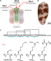
Binding site of μO§-GVIIJ on the channel and its peptide sequence. A, binding sites of VGSC-inhibiting conotoxins. Pore-blocking μ-conotoxins bind deep within the Na+ conductance pore and compete with tetrodotoxin for neurotoxin binding site 1. Site 1 is located near the bottom of the reentrant loops in all four domains (domains I–IV), but only one site of interaction is shown in this figure. In contrast, μO-conotoxins MrVIA/B bind to the voltage sensor of domain II at neurotoxin binding site 4 (3), whereas μO§-GVIIJ, whose mechanism of block remains to be determined, binds to the loop in the pore module of domain II at site 8 (4). B, photograph of the shell of the fish-hunting cone snail C. geographus. C, primary sequence of μO§-GVIIJ, displaying the previously determined disulfide framework (4). Intercysteine loops 1–4 are underlined. X represents modifications at position 24 used in the structural and pharmacological studies described in this report; their structures are shown at the bottom.
FIGURE 2.
FIGURE 2.
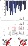
Characterization of GVIIJ[C24S] by NMR spectroscopy. A, temperature dependence experiments identified several residues that exhibited amide proton chemical shifts of magnitude < 4 ppb/K, including Trp-2, Arg-14, Cys-18, Gly-20, Phe-21, Asp-23, Thr-26, Thr-28, Cys-29, and Lys-30. These shifts suggest partial protection of the residues from solvent. B, amide exchange experiments demonstrated that Cys-10, Lys-12, Arg-14, Leu-15, Cys-29, Phe-21, Lys-30, and Ser-33 exchanged slowly in 100% 2H2O at 25 °C. These amide protons were either inaccessible to solvent through their location in the folded peptide or were protected from exchange by interactions such as hydrogen bonds. Amide and aromatic regions of one-dimensional 1H NMR spectra in 100% 2H2O at 25 °C between 0 and 14 h are shown. The data were acquired using a Bruker DRX-600 spectrometer. Rapidly exchanging amide protons (0–1 h) are listed in black, medium exchange protons (1–8 h) are in pink, and slowly exchanging amide protons (>8 h) are in red. C, locations of rapidly exchanging amide protons mapped onto the structure of GVIIJ[C24S] (see below). Fast (white), medium (pink), and slow (red) exchanging amide protons are shown on the closest to average structure of GVIIJ[C24S]. The structure is rotated by 180° about the y axis to show labile protons on the opposite face of the peptide.
FIGURE 3.
FIGURE 3.
Deviation of backbone amide (HN) and Cα proton (Hα) chemical shifts from random coil values at 25 °C (28). A, HN chemical shift deviations for GVIIJ[C24S] (red) and GVIIJSSEA (blue). B, Hα chemical shift deviations for GVIIJ[C24S] (red) and GVIIJSSEA (blue). O is hydroxyproline. X is either serine (GVIIJ[C24S]) or S-((2-aminoethyl)thio)cysteine (GVIIJSSEA).
FIGURE 4.
FIGURE 4.
Structure of the μO§-GVIIJ analog GVIIJ[C24S]. A, family of 20 final structures for GVIIJ[C24S], superimposed on backbone heavy atoms of residues 2–32. Disulfide bonds are shown in orange according to the previously established connectivity: Cys-3–Cys-18, Cys-10–Cys-22, and Cys-17–Cys-29. B, ribbon representation of the closest to average structure with calculated β-sheets at residues 22–23 (β1) and 28–29 (β2). Position of Ser-24 (in loop 4) is indicated. In the panel on the left, residues 8–10 are located in the looped region adjacent to the β2-strand of GVIIJ[C24S]. C, surface representation of GVIIJ[C24S], with basic residues colored light and dark blue (Arg and Lys, respectively); acidic residues (Asp) colored red; Cys residues colored orange; aromatic residues (Trp and Tyr) colored purple; and all others shown in white. Depictions rotated by 180° about the y axis are also shown.
FIGURE 5.
FIGURE 5.
Characterization of the final 20 structures of GVIIJ[C24S]. A, sequential and medium-range NOE connectivities for GVIIJ[C24S] (298 K, pH 3.2) according to CYANA structure calculation. Intensities of dNN, dαN, and dβN are proportional to the height of the bars. Measurable 3JHNHα are noted as follows: 3JHNHα < 6 Hz (↓),3JHNHα 6–8 Hz (○), and 3JHNHα > 8 Hz (↑). Amide proton exchange rates for residues 1–35 are designated as follows: white box, fast exchange rate (0–1 h); gray box, medium exchange rate (1–8 h); and black box, slow exchange rate (>8 h). Regions containing elements of secondary structure (β-strands between Cys-22–Asp-23 and Thr-28–Cys-29) are indicated. B, CYANA output showing the number of long-range (ij ≥ 6), short-range (2 ≤ ij ≤ 5) and intraresidue NOE restraints used in the calculation of the final structure. C, minimal backbone structure of the ensemble of 20 structures of GVIIJ[C24S] showing disordered N and C termini (red) and the dynamic loop 2 region (Cys-11–Tyr-16) of the peptide (purple). D–F, angular order parameters (S) for backbone (φ, ψ) and sidechain (χ1) dihedral angles.
FIGURE 6.
FIGURE 6.
Structure-activity relationship studies of residues in the Cys-24-containing loop 4. A, the closest to average backbone structure of GVIIJ[C24S] is stabilized by numerous H-bonds between backbone atoms, as well as by side chain to backbone interactions between the residues in this loop (Phe-21–Thr-28). A type I β-turn between two antiparallel β-strands is stabilized by interactions between residues in this turn (Asp-23–Thr-26). B, HPLC elution profiles of GVIIJSSEA analogs following folding. Conventional folding of Ala-replacement mutants of Phe-21, Asp-23, Thr-26, and Thr-28 did not yield a single, major folding species (black trace); however, folding in the presence of l-cystine (red trace) resulted in a major product (red asterisk), although with significantly lower yields compared with other mutants (black asterisks). The [D5K] analog was included to show that charge reversal did not affect accumulation of a major product when the substitution was made to residues spatially distinct from loop 4. C, comparison of NaV1.2 blockade by GVIIJSSEA mutants in the Cys-24 containing loop. The data are from Table 3. Analogs constructed on the GVIIJSSC background are denoted by a red asterisk. Nearly all analogs were functionally equipotent with the unmodified peptide, suggesting that residues in this region are important for stability, but not activity. One exception to this was the [T28A] analog, which exhibited a Kd value of 8.60 ± 2.30 nm, significantly higher (p value = 0.0212) than that of unmodified peptide. Selection of some amino acid replacements in this loop was based on modest sequence homology between loop 4 of μO§-GVIIJ and rat or human NaVβ2 and β4-subunits, whose partial homologous sequences are shown in the inset.
FIGURE 7.
FIGURE 7.
NaV1.2 block by μO§-GVIIJ analogs. A, comparison of NaV1.2 blockade by 3.3 μm (filled circles) and 33 μm (open circles) GVIIJ[C24S]. Block was rapidly reversible at all concentrations tested. Responses did not completely return to baseline following washout of high concentrations of peptide; studies are underway to better understand this phenomenon. The inset shows the current traces before (black) and after (red) application of 33 μm GVIIJ[C24S]. B, dose-response curve for GVIIJ[C24S]. The data points are means ± S.E. (n ≥ 3 oocytes). The solid line is the best fit curve to the equation % peak INa blocked = Ymax/(1 + (IC50/[peptide])), where Ymax, the extrapolated level of block at saturating peptide concentrations, was 82.8% (with 95% CI of 77.5–88.10), and the IC50 was 4.8 μm (with 95% CI of 3.7–6.2 μm). C, GVIIJSSEA (1 μm) rapidly blocked ∼70% of the sodium current. In contrast to the [C24S] analog, this analog exhibited nearly irreversible block of NaV1.2 following washout, which is attributed to tethering to site 8 of the channel (4, 5). D–F, substitution of critical amino acids Lys-12, Arg-14, and Tyr-16 resulted in analogs with lower kobs for the inhibition of NaV1.2 (see also Table 3). The red bar represents when the oocyte was exposed to peptide. The inset in each panel shows the current traces before (black) and during (red) exposure to the indicated peptides.
FIGURE 8.
FIGURE 8.
Block of NaV1. 4 by μ-PIIIA[R14Q], μ-GIIIA, μO§-GVIIJ, or (μO§-GVIIJ)2, alone or in binary combinations. Peak INa of voltage clamped oocytes expressing rat NaV1.4 were monitored as described under “Experimental Procedures.” The bars in representative time courses of block indicate when peptides were present in the (static) bath. The values are means ± S.D. or 95% CI (n ≥ 5 oocytes). A, concentration-response curve of μ-PIIIA[R14Q]; solid line is best fit curve to the binding equation provided in Fig. 7; IC50 = 0.25 μm (95% CI of 0.19–0.33 μm) and percentage block at saturating [peptide] was 73 ± 3.5% (dashed line) (i.e. residual current, rINa, was 27%). Inset, representative time course of block of INa by 33 μm μ-PIIIA[R14Q] and recovery following peptide washout; average value of koff was 1.2 ± 0.32 min−1. B, as in the inset of A, except that after the block by μ-PIIIA[R14Q] had reached steady state, the bath was supplemented with μ-GIIIA (final concentration, 10 μm) to observe its block of rINa, and then both peptides were washed out. Inset, time course of block by 10 μm μ-GIIIA alone. Note, μ-GIIIA blocked the rINa much more slowly than the control INa illustrated in the inset, which indicates that μ-PIIIA[R14Q] and μ-GIIIA competed for the same site to block the channel. C, as in B, except the bath was supplemented with μO-GVIIJSSG (final concentration, 3.3 μm) to observe its block of rINa. Inset, time course of block by 3.3 μm μO-GVIIJSSG alone. Note, μO-GVIIJSSG blocked rINa essentially as fast as it did control INa illustrated in the inset, which indicates that μ-PIIIA[R14Q] and μO§-GVIIJ did not compete with each other to block the channel. D, as in B, except the bath was supplemented with 33 μm (μO§-GVIIJ)2 to observe its block of rINa. Inset, time course of block by 33 μm (μO§-GVIIJ)2 alone. Although not evident by visual inspection, (μO-GVIIJ)2 blocked rINa slightly more slowly than it did control INa (see Table 4 for average kobs values).
FIGURE 9.
FIGURE 9.
Structural comparison of the GVIIJ[C24S] backbone with those of other ICK motif-containing peptides. The backbone of the closest to average structure of GVIIJ[C24S] (blue) was aligned with the backbones of δ-conotoxin EVIA from Conus ermenius (Protein Data Bank code 1G1P, A), Ptu1 toxin from the assassin beetle (Protein Data Bank code 1I26, B), purotoxin-1 from the burrowing wolf spider (Protein Data Bank code 2KGU, C) and vesutoxin from the Australian Blue Mountain funnel web spider (Protein Data Bank code 1AHX, D). Structural alignments were made using DaliLite version 3 (37). The ion channel modulating toxins shown in this figure were selected based on the following criteria: sequence homology ≥ 30%, similar size (30–36 amino acid residues), and a Z score ≥ 2.0.
FIGURE 10.
FIGURE 10.
“Functionally bipartite” mechanism of action for μO§-GVIIJ. A, locations of amino acid residues important for on rates (kon) of μO§-GVIIJ against NaV1.2. Residues with the slowest kon values are colored red. B, location of residue 24 (magenta), which is responsible for the covalent interactions with a free cysteine on the channel (Cys-910). Replacement of Cys-24 with serine prevented disulfide bridge formation between the peptide and the channel and led to rapid reversibility upon washout, indicating that the identity of the residue in position 24 is critical for koff. C, backbone representation of GVIIJ[C24S], which shows the locations of residues deemed particularly important for the activity of μO§-GVIIJ. Residues important for the on rate (Lys-12, Arg-14, and Tyr-16) of the peptide are located in loop 2 (red), whereas that affecting the off rate is located within the β-turn of loop 4 (magenta).

Similar articles

Cited by

References

    1. Robinson S. D., and Norton R. S. (2014) Conotoxin gene superfamilies. Mar. Drugs 12, 6058–6101 - PMC - PubMed
    1. Fiedler B., Zhang M. M., Buczek O., Azam L., Bulaj G., Norton R. S., Olivera B. M., and Yoshikami D. (2008) Specificity, affinity and efficacy of ι-conotoxin RxIA, an agonist of voltage-gated sodium channels NaV1.2, 1.6 and 1.7. Biochem. Pharmacol. 75, 2334–2344 - PMC - PubMed
    1. Leipold E., DeBie H., Zorn S., Borges A., Olivera B. M., Terlau H., and Heinemann S. H. (2007) μO-conotoxins inhibit NaV channels by interfering with their voltage sensors in domain-2. Channels 1, 253–262 - PubMed
    1. Gajewiak J., Azam L., Imperial J., Walewska A., Green B. R., Bandyopadhyay P. K., Raghuraman S., Ueberheide B., Bern M., Zhou H. M., Minassian N. A., Hagan R. H., Flinspach M., Liu Y., Bulaj G., Wickenden A. D., Olivera B. M., Yoshikami D., and Zhang M. M. (2014) A disulfide tether stabilizes the block of sodium channels by the conotoxin μO§-GVIIJ. Proc. Natl. Acad. Sci. U.S.A. 111, 2758–2763 - PMC - PubMed
    1. Zhang M. M., Gajewiak J., Azam L., Bulaj G., Olivera B. M., and Yoshikami D. (2015) Probing the redox states of sodium channel cysteines at the binding site of μO§-conotoxin GVIIJ. Biochemistry 54, 3911–3920 - PubMed

Publication types

MeSH terms

LinkOut - more resources