. 2016 Mar 25;291(13):7097-106.
doi: 10.1074/jbc.M115.680611. Epub 2016 Jan 27.

Scorpion Potassium Channel-blocking Defensin Highlights a Functional Link with Neurotoxin

Affiliations

Scorpion Potassium Channel-blocking Defensin Highlights a Functional Link with Neurotoxin

Lanxia Meng et al. J Biol Chem. .

Abstract

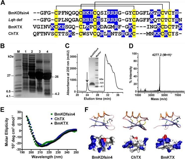
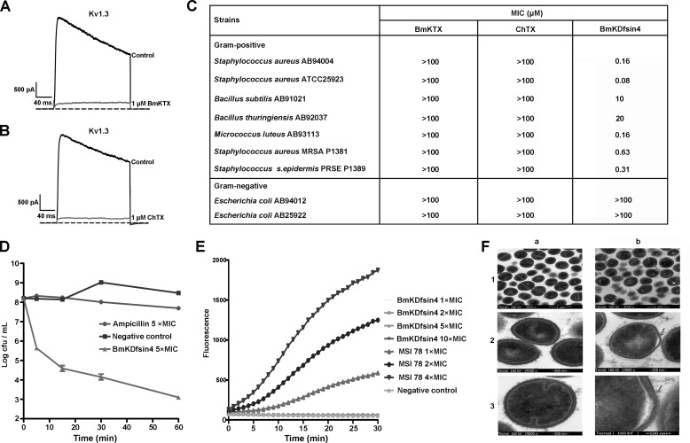
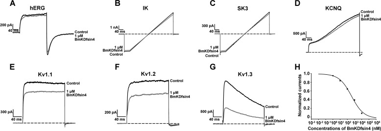
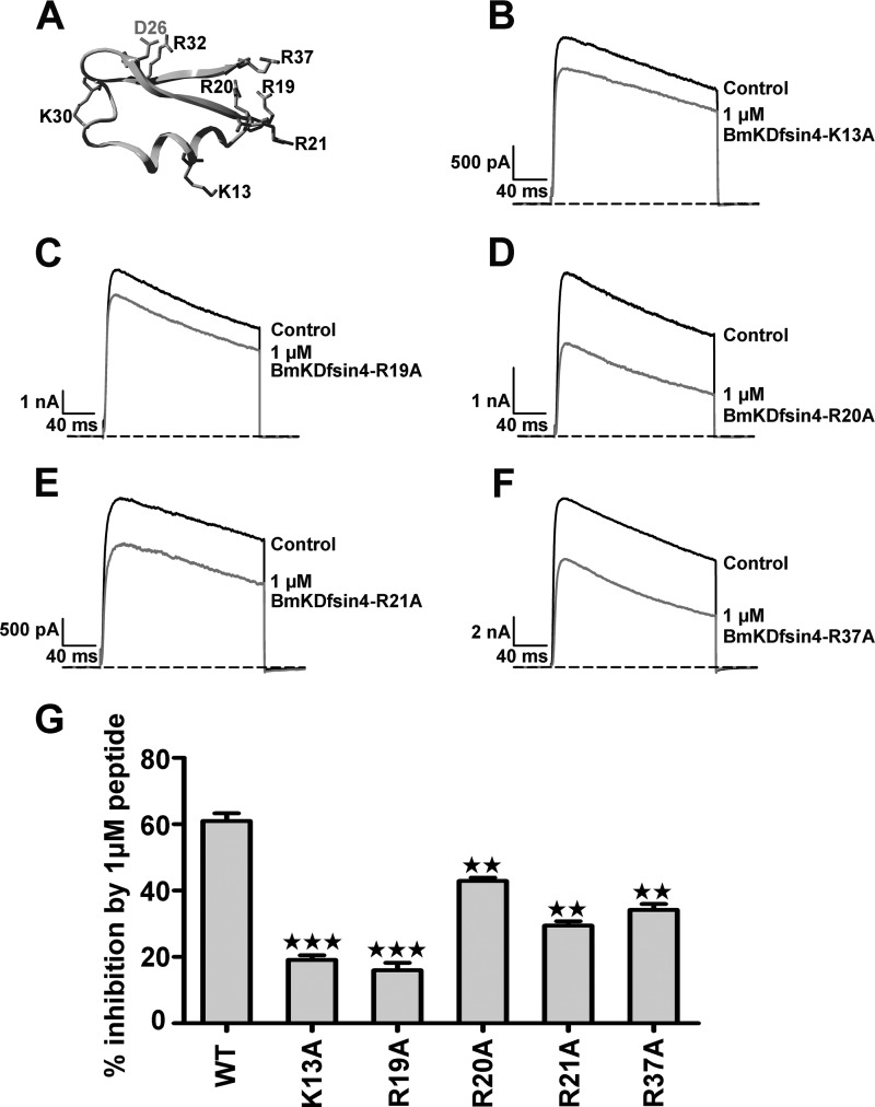
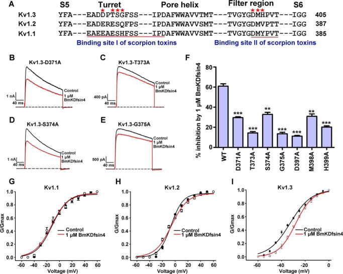
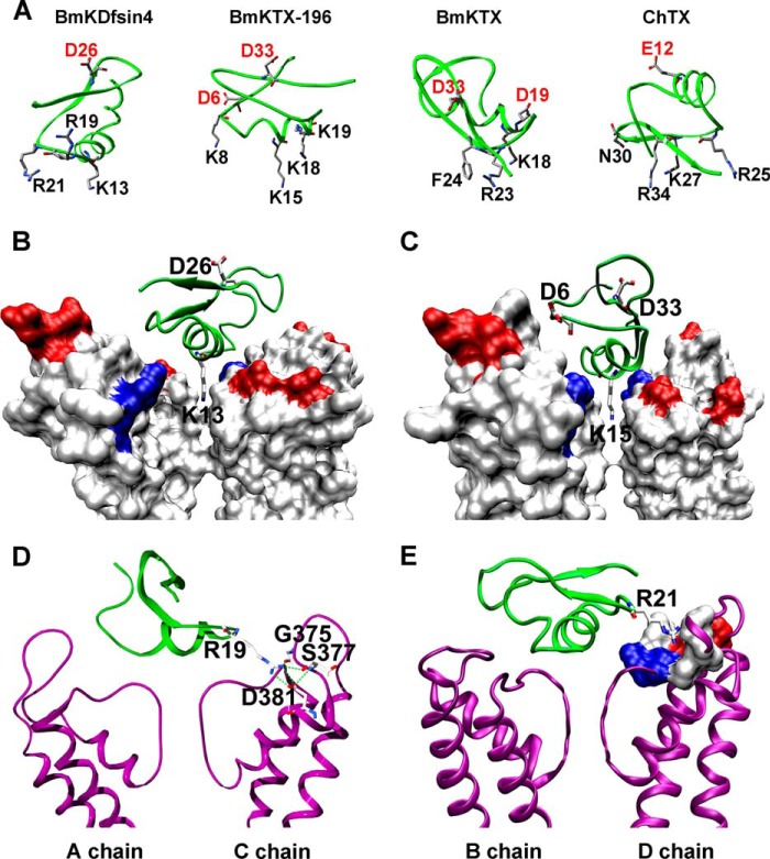
The structural similarity between defensins and scorpion neurotoxins suggests that they might have evolved from a common ancestor. However, there is no direct experimental evidence demonstrating a functional link between scorpion neurotoxins and defensins. The scorpion defensin BmKDfsin4 from Mesobuthus martensiiKarsch contains 37 amino acid residues and a conserved cystine-stabilized α/β structural fold. The recombinant BmKDfsin4, a classical defensin, has been found to have inhibitory activity against Gram-positive bacteria such as Staphylococcus aureus, Bacillus subtilis, and Micrococcus luteusas well as methicillin-resistant Staphylococcus aureus Interestingly, electrophysiological experiments showed that BmKDfsin4,like scorpion potassium channel neurotoxins, could effectively inhibit Kv1.1, Kv1.2, and Kv1.3 channel currents, and its IC50value for the Kv1.3 channel was 510.2 nm Similar to the structure-function relationships of classical scorpion potassium channel-blocking toxins, basic residues (Lys-13 and Arg-19) of BmKDfsin4 play critical roles in peptide-Kv1.3 channel interactions. Furthermore, mutagenesis and electrophysiological experiments demonstrated that the channel extracellular pore region is the binding site of BmKDfsin4, indicating that BmKDfsin4 adopts the same mechanism for blocking potassium channel currents as classical scorpion toxins. Taken together, our work identifies scorpion BmKDfsin4 as the first invertebrate defensin to block potassium channels. These findings not only demonstrate that defensins from invertebrate animals are a novel type of potassium channel blockers but also provide evidence of a functional link between defensins and neurotoxins.

Keywords: bacteria; defensin; neurotoxin; potassium channel; structure-function.


Figures

FIGURE 1.
FIGURE 1.
Sequence alignment, production, and structural analysis of BmKDfsin4. A, multiple amino acid sequence alignment of BmKDfsin4, Lqh def (a scorpion defensin from L. quinquestriatus), ChTX (a toxin from L. quinquestriatus hebraeus), and BmKTX (a native toxin from M. martensii Karsch). Cysteines are shadowed in yellow, and basic residues are shown in blue. B, Tricine SDS-PAGE analysis of the expression of the GST-BmKDfsin4 fusion protein and split products. Lane M, molecular mass markers; lane 1, total cell-free extract of E. coli carrying pGEX-6p-1-BmKDfsin4 without isopropyl 1-thio-β-d-galactopyranoside induction; lane 2, total cell-free extract of E. coli carrying pGEX-6p-1-BmKDfsin4 induced with 0.1 mm isopropyl 1-thio-β-d-galactopyranoside; lane 3, the purified GST-BmKDfsin4 fusion protein after desalting and concentration; lane 4, the fusion protein cleaved by enterokinase. C, the HPLC profile of the GST-BmKDfsin4 fusion protein cleaved by enterokinase. The fraction containing BmKDfsin4 is indicated by the arrow and was analyzed by Tricine SDS-PAGE. D, mass spectrum of the BmKDfsin4 peptide measured by MALDI-TOF/MS. The measured value is 4277.2 Da, and the calculated value is 4276.9 Da. E, the circular dichroism spectra of BmKDfsin4, measured at 25 °C from 195–250 nm. Data were collected at 1-nm intervals with a continuous scan rate of 200 nm/min. F, three-dimensional structures of BmKDfsin4, ChTX, and BmKTX (top row). The disulfide bonds are shown. Also shown are space-filling molecular surfaces of BmKDfsin4, ChTX, and BmKTX (bottom row) showing the locations of different types of amino acids (blue, basic residues; red, acidic residues).
FIGURE 2.
FIGURE 2.
The antibacterial activity and mechanism of BmKDfsin4. A, representative current traces in the absence (control) or presence of 1 μm BmKTX on the currents of the Kv1.3 channel. 1 μm BmKTX blocked 94.9% ± 1.1% of Kv1.3 channel currents. B, representative current traces in the absence (control) or presence of 1 μm ChTX on the currents of the Kv1.3 channel. 1 μm ChTX blocks 93.0% ± 3.1% of Kv1.3 channel currents. Each experiment of channel current blockage was performed at least three times (n ≥ 3). C, MICs of BmKTX, ChTX, and BmKDfsin4 against bacteria. Every concentration of peptides was set three parallel duplicates, and only the concentration for all three parallel samples without bacterial growth was used as the MIC of the peptide against bacteria. MIC determination of peptides was repeated three times (n = 3), and the determined MIC values were the same. D, killing kinetics of BmKDfsin4 against S. aureus AB94004. The bacteria were treated with BmKDfsin4 or ampicillin at 5 × MIC. The experiment was repeated with similar results. E, the effect of BmKDfsin4 on bacterial membrane integrity. BmKDfsin4 was used at 1 × MIC, 2 × MIC, 5 × MIC, and 10 × MIC. The negative and positive controls were 0.9% saline and MSI-78 (an amphipathic α-helical peptide with an antibacterial mechanism of disrupting the membrane), respectively. The MIC of MSI-78 against S. aureus AB94004 was 3.53 μm. The experiment was repeated with similar results. F, transmission electron microscopy observation of S. aureus AB94004 in the absence or presence of BmKDfsin4. a1–3, negative control. b1–3, treatment with BmKDfsin4 at a concentration of 10 × MIC for 30 min. Cells showing plasmolysis are indicated by arrows. Each experiment was performed in duplicate (n = 3).
FIGURE 3.
FIGURE 3.
The BmKDfsin4 interaction with potassium channels. A–G, representative current traces in the absence (control) or presence of 1 μm BmKDfsin4 on the currents from the potassium channels hERG, IK, SK3, KCNQ, Kv1.1, Kv1.2, and Kv1.3. At 1 μm, BmKDfsin4 blocked 11.6% ± 1.2% of ERG, 3.9% ± 0.3% of IK, 7.0% ± 0.6% of SK3, 5.9% ± 0.5% of KCNQ, 25.2% ± 1.9% of Kv1.1, 30.5% ± 1.1% of Kv1.2, and 61.0% ± 1.6% of Kv1.3 currents. The Kv1.1 channel is from mice, and the other channels are from humans. The black and red lines represent the currents measured in the absence (control) and presence of BmKDfsin4, respectively. H, average normalized hKv1.3 channel current inhibition by various concentrations of BmKDfsin4. The IC50 was 510.2 ± 161.2 nm. Each channel was tested at least three times (n ≥ 3). The results are shown as mean ± S.E.
FIGURE 4.
FIGURE 4.
The effect of basic residues on the binding affinity of BmKDfsin4. A, the basic residue distribution in the three-dimensional structure of BmKDfsin4. B–F, at 1 μm, BmKDfsin4-K13A, R19A, R20A, R21A, and R37A cause inhibitions of 19.1% ± 1.4%, 16.0% ± 2.3%, 42.9% ± 1.0%, 29.4% ± 1.4%, and 34.2% ± 1.8%, respectively. The black and red lines represent the currents measured in the absence (control) and presence of BmKDfsin4, respectively. G, average inhibition of Kv1.3 channel currents by 1 μm wild-type or mutant BmKDfsin4. The control current amplitude in each experiment was fixed as 1 for the normalized currents, and the inhibition rates were compared. Each channel was tested at least three times (n ≥ 3). The results are shown as mean ± S.E. ★★★, p < 0.001; ★★, p < 0.01).
FIGURE 5.
FIGURE 5.
The Kv1.3 channel outer vestibule is responsible for BmKDfsin4 binding. A, multiple sequence alignment of the outer vestibules of the hKv1.3, hKv1.2, and mKv1.1 channels. B–E, representative current traces of the Kv1.3 channel with mutations in the pore region in response to 1 μm BmKDfsin4. At 1 μm, BmKDfsin4 inhibited potassium currents as follows: 29.6% ± 0.8% for the Kv1.3-D371A channel, 14.4% ± 1.4% for the Kv1.3-T373A channel, 32.8% ± 2.0% for the Kv1.3-S374A channel, and 13.5% ± 1.5% for the Kv1.3-G375A channel. The black and red lines represent the currents measured in the absence (control) and presence of BmKDfsin4, respectively. F, average inhibition of wild-type and mutant Kv1.3 channel currents by 1 μm BmKDfsin4. The control current amplitude in each experiment was fixed as 1 for the normalized currents, and the inhibition rates were compared. Each channel was tested at least three times (n ≥ 3). The results are shown as mean ± S.E. (***, p < 0.001; **, p < 0.01. G–I, the conductance-voltage curves from the peak currents were plotted for Kv1.1, Kv1.2, and Kv1.3 in the absence and presence of 1 μm BmKDfsin4. No significant G-V curve shift was observed. The detailed ΔV50 values before and after BmKDfsin4 interacting with Kv1.1, Kv1.2, and Kv1.3 channels were −0.61, 0.82, and 4.62 mV, respectively.
FIGURE 6.
FIGURE 6.
The interaction model between BmKDfsin4 and the tetrameric Kv1. 3 channel. A, the distribution of acidic residues and main functional residues of BmKDfsin4, BmKTX-196, BmKTX, and ChTX. B, Lys-13 of BmKDfsin4 interacts with residues around the selectivity filter of the Kv1.3 channel. C, Lys-15 of BmKTX196 interacts with residues around the selectivity filter of the Kv1.3 channel (22) D, Arg-19 of BmKDfsin4 interacts with the Kv1.3 channel through hydrogen bonds and salt bridges. E, Arg-21 of BmKDfsin4 interacts with polar and nonpolar residues in the Kv1.3 channel.

References

    1. Dunlop J. A., Kamenz C., and Scholtz G. (2007) Reinterpreting the morphology of the Jurassic scorpion Liassoscorpionides. Arthropod Struct. Dev. 36, 245–252 - PubMed
    1. Fry B. G., Roelants K., Champagne D. E., Scheib H., Tyndall J. D., King G. F., Nevalainen T. J., Norman J. A., Lewis R. J., Norton R. S., Renjifo C., and de la Vega R. C. (2009) The toxicogenomic multiverse: convergent recruitment of proteins into animal venoms. Annu. Rev. Genomics Hum. Genet. 10, 483–511 - PubMed
    1. Batista C. V., D'Suze G., Gómez-Lagunas F., Zamudio F. Z., Encarnación S., Sevcik C., and Possani L. D. (2006) Proteomic analysis of Tityus discrepans scorpion venom and amino acid sequence of novel toxins. Proteomics 6, 3718–3727 - PubMed
    1. de Oliveira U. C., Candido D. M., Coronado Dorce V. A., and Junqueira-de-Azevedo I. L. (2015) The transcriptome recipe for the venom cocktail of Tityus bahiensis scorpion. Toxicon 95, 52–61 - PubMed
    1. Xu X., Duan Z., Di Z., He Y., Li J., Li Z., Xie C., Zeng X., Cao Z., Wu Y., Liang S., and Li W. (2014) Proteomic analysis of the venom from the scorpion Mesobuthus martensii. J. Proteomics 106, 162–180 - PubMed

Publication types

MeSH terms