

Extrathyroidal Calcitonin Secreting Tumors: Pancreatic Neuroendocrine Tumors in Patients With Multinodular Goiter: Two Case Reports
- PMID: 26817871
- PMCID: PMC4998245
- DOI: 10.1097/MD.0000000000002419
Extrathyroidal Calcitonin Secreting Tumors: Pancreatic Neuroendocrine Tumors in Patients With Multinodular Goiter: Two Case Reports
Abstract
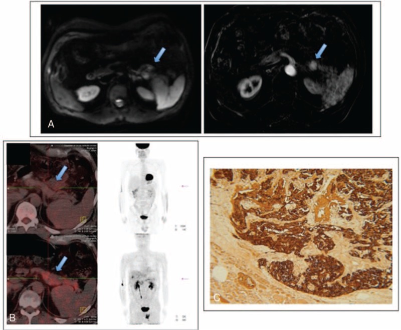
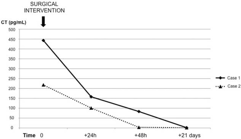
Calcitonin is the hallmark of medullary thyroid carcinoma. However, extrathyroidal neuroendocrine tumors can also release calcitonin.We report 2 cases of calcitonin-secreting pancreatic tumors found in asymptomatic patients with thyroid nodules referred to our center within 11 months.Case 1: A man initially referred for thyroid nodule characterization was found to have hypercalcitoninemia (>200 pg/mL) during non-neoplastic fine-needle aspiration.Case 2: A woman evaluated for liver metastasis was found to have hypercalcitoninemia and multinodular goiter.Our research emphasizes that marked hypercalcitoninemia in the presence of thyroid nodules is not necessarily due to medullary thyroid carcinoma; awareness of this could avoid unnecessary thyroidectomy. The lack of specific symptoms related to hypercalcitoninemia may be the reason that the prevalence of calcitonin-secreting pancreatic tumors is underestimated.
Conflict of interest statement
The authors have no conflicts of interest to disclose.
Figures
References
-
- Cheung K, Roman SA, Wang TS, et al. Calcitonin measurement in evaluation of thyroid nodules in the United States: a cost-effectiveness and decision analysis. J Clin Endocrinol Metab 2008; 93:2173–2180. - PubMed
-
- Cvijovic G, Micic D, Kendereski A, et al. Ectopic calcitonin secretion in a woman with large cell neuroendocrine lung carcinoma. Hormones (Athens) 2013; 12:584–590. - PubMed
-
- Schwartz KE, Wolfsen AR, Forster B, et al. Calcitonin in nonthyroidal cancer. J Clin Endocrinol Metab 1979; 49:438–444. - PubMed
-
- Schneider R, Waldmann J, Swaid Z, et al. Calcitonin-secreting pancreatic endocrine tumors: systematic analysis of a rare tumor entity. Pancreas 2011; 40:213–221. - PubMed
-
- Eriksson B, Arnberg H, Lindgren PG, et al. Neuroendocrine pancreatic tumours: clinical presentation, biochemical and histopathological findings in 84 patients. J Intern Med 1990; 228:103–113. - PubMed
Publication types
MeSH terms
Substances
LinkOut - more resources
Full Text Sources
Other Literature Sources
Medical
