. 2016 Jan;95(3):e2524.
doi: 10.1097/MD.0000000000002524.

Selective Retina Therapy in Patients With Chronic Central Serous Chorioretinopathy: A Pilot Study

Affiliations

Selective Retina Therapy in Patients With Chronic Central Serous Chorioretinopathy: A Pilot Study

Seungbum Kang et al. Medicine (Baltimore). 2016 Jan.

Erratum in

Abstract

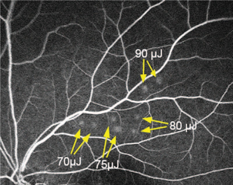
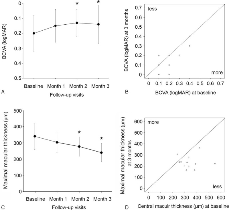
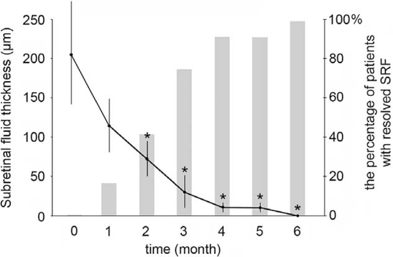
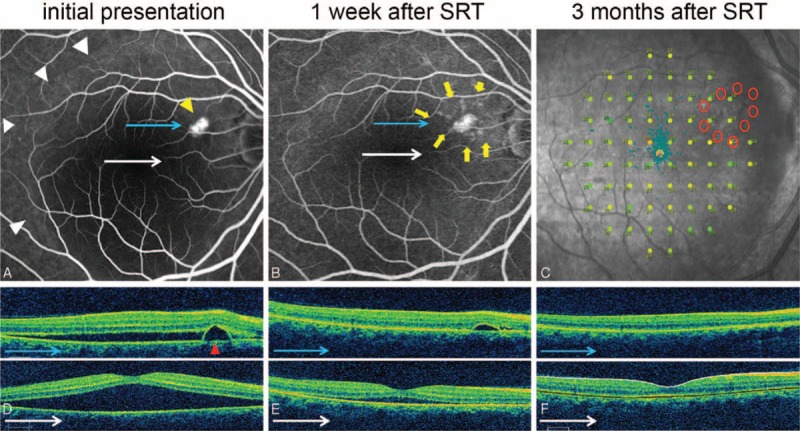
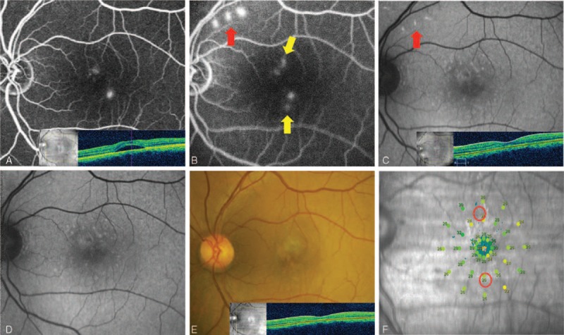
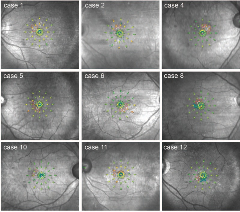
We evaluated visual outcomes, changes of maximum macular thickness (MMT) and subretinal fluid (SRF), and safety in patients with chronic central serous chorioretinopathy (CSC) after treatment with selective retina therapy (SRT). Retrospective cohort study of patients with chronic CSC presenting to a university-based hospital from January 2014 through January 2015 was conducted. A total of 12 eyes of 12 patients with chronic CSC lasting for at least 3 months was recruited. The follow-up period ranged from 3 to 12 months. Following evaluation of test spots at temporal arcades, SRT (Q-switched neodymium-doped yttrium lithium fluoride [Nd:YLF] laser; wavelength, 527 nm, pulse duration, 1.7 microsececond) was applied to the surrounding areas of leakage observed on fluorescein angiogram and/or pigment epithelial detachment (PED). Changes in best-correct visual acuity (BCVA), MMT, and SRF and macular sensitivity (MS) by microperimetry (MP) were evaluated. Eyes received treatment in a mean of 3.83 spots at the pulse energy of 65 to 90 μJ. Mean BCVA (logMAR) improved from 0.23 ± 0.12 at baseline to 0.14 ± 0.13 at 3 months. MMT decreased from 341.4 ± 85.5 μm at baseline to 236.0 ± 57.9 μm at 3 months. SRF completely resolved in 75% (9 eyes) at 3 months. Large PEDs (2 eyes) were flattened at 3 months. Retreatment was performed in 4 eyes. MP showed no evidence of scotoma around SRT-treated lesions. SRT treatment targeting the surrounding area of leakage point showed favorable visual and structural outcomes in chronic CSC patients without the risk of scotoma.


Conflict of interest statement

The authors have no conflicts of interest to disclose.

Figures

FIGURE 1
FIGURE 1
Representative image of fundus fluorescein angiography showing 8 test spots. Each pulse energy with 70, 75, 80, and 90 μJ was tested in duplicate.
FIGURE 2
FIGURE 2
Scatter plot of reflectometry and optoacustic dosimetry demonstrating the value of each test spot. In this study, 68 of 73 test spots showed hyperfluorescence on fundus fluorescein angiography (FFA) and no overtreatment was observed in any of the selective retina therapy (SRT) spots. Spots without cell damage (FFA invisible; red circles) and selective damaged retinal pigment epithelium cells (FFA visible; green circles) are shown. The energy of treatment spots was chosen among FFA-positive spots within the range of the blue rectangle.
FIGURE 3
FIGURE 3
(A) Change of best-correct visual acuity (BCVA) (logMAR) in selective retina therapy (SRT)-treated patients with chronic central serous chorioretinopathy (CSC). Error bars represent standard deviation (SD). The difference of BCVA (logMAR) was statistically significant at 2 and 3 months after SRT. P < 0.05. (B) Scatter plot of BCVA (logMAR) of patients at baseline and at 3 months. (C) Change of maximum macular thickness (MMT) measured by optical coherence tomography (OCT) in SRT-treated patients with chronic CSC. Error bars represent SD. The difference of MMT was statistically significant at 2 and 3 months after SRT. P < 0.05. (D) Scatter plot of MMT of patients at baseline and at 3 months.
FIGURE 4
FIGURE 4
Changes of subretinal fluid (SRF) thickness (left axis) and the percentage of patient with completely resolved SRF (right axis) in SRT-treated patients with chronic central serous chorioretinopathy (CSC). Error bars represent SD. P < 0.05.
FIGURE 5
FIGURE 5
Case 7: A 46-year-old woman presented with a 3-month history of distortion of central vision in the right eye. Her right best-correct visual acuity (BCVA) was 20/32. Selective retina therapy (SRT) was applied to the area surrounding the pigment epithelial detachment (PED). Subretinal fluid (SRF) resolved markedly and PED was flattened prominently in 1 week. Both SRF and PED disappeared within 3 months. The BCVA improved to 20/20 in her right eye. (A) Large SRF (white arrowheads) with PED (yellow arrowhead) was observed on fundus fluorescein angiography (FFA) at baseline. (B) FFA demonstrated nine SRT laser spots (yellow arrows) surrounding PED at 1 week after SRT treatment. (C) Microperimetry (MP) performed 4 months after SRT treatment showed no significant decrease or scotoma change at SRT-treated regions. (D) Baseline optical coherence tomography (OCT) showed PED (red arrowhead) and large SRF. (E) PED and SRF were rapidly decreased at 1 week after SRT treatment. (F) The SRF and PED were completely resolved at 3 months after SRT.
FIGURE 6
FIGURE 6
Case 9: A 41-year-old man presented with a 4-month history of central blur in the right eye. The best-correct visual acuity (BCVA) was 20/25. Previous 3 times of intravitreal injection of bevacizumab did not show any improvement. Selective retina therapy (SRT) was applied to the area surrounding the leaking point. Although both subretinal fluid (SRF) and pigment epithelial detachment (PED) disappeared completely, his BCVA remained unchanged. (A) Active leaking at PED was observed on fundus fluorescein angiography (FFA). Six arch-shaped SRT treatment spots (6 red circles) were applied around the leaking point. (B) No active leakage was found at PED (yellow arrow) on FFA. (C) Microperimetry (MP) demonstrated no scotoma on macular sensitivity. (D) Optical coherence tomography (OCT) showed PED (red arrowhead) and SRF at macular region at baseline. (E) OCT showed rapid resolution of SRF and PED 2 weeks after SRT treatment. (F) SRF completely disappeared and PED was nearly flattened at 3 months after SRT treatment.
FIGURE 7
FIGURE 7
Case 3: A 45-year-old man presented with a 12-month history of central serous chorioretinopathy (CSC) in the left eye. His left best-correct visual acuity (BCVA) was 20/50. He had been treated with intravitreal injection of bevacizumab about 4 months previously. Selective retina therapy (SRT) was applied to the area of focal juxtafoveal angiographic leakages. His persistent subretinal fluid (SRF) completely resolved within 1 month. The BCVA remained unchanged. (A) Two leaking points on fundus fluorescein angiography (FFA) and SRF on optical coherence tomography (OCT) were observed at initial presentation. (B) Two SRT laser spots near 2 leaking points (yellow arrow) and 3 test spots (red arrow) were shown on FFA 30 minutes after SRT treatment. (C) Fundus autofluorescence (FAF) showed hyperautofluorescence at test spots at 1 month. (D) Hyperautofluorescence at test spots nearly disappeared at 3 months. (E) No scar was seen at SRT-treated sites. Complete resolution of SRF was shown on OCT 3 months after SRT. (F) Microperimetry (MP) showed no scotoma at 4 months after SRT.
FIGURE 8
FIGURE 8
Microperimetry (MP) data were obtained after the complete resolution of subretinal fluid in selective retina therapy (SRT)-treated 12 patients. In all cases, there was no detectable scotoma in any of the SRT-treated lesions. Red circles indicate the SRT-irradiated points. White circles indicate leak points shown in fundus fluorescein angiography.

References

    1. Wang M, Munch IC, Hasler PW, et al. Central serous chorioretinopathy. Acta Ophthalmol 2008; 86:126–145. - PubMed
    1. Spitznas M. Pathogenesis of central serous retinopathy: a new working hypothesis. Graefes Arch Clin Exp Ophthalmol 1986; 224:321–324. - PubMed
    1. Spaide RF, Goldbaum M, Wong DW, et al. Serous detachment of the retina. Retina 2003; 23:820–846. - PubMed
    1. Liew G, Quin G, Gillies M, et al. Central serous chorioretinopathy: a review of epidemiology and pathophysiology. Clin Exp Ophthalmol 2013; 41:201–214. - PubMed
    1. Cardillo Piccolino F, Eandi CM, Ventre L, et al. Photodynamic therapy for chronic central serous chorioretinopathy. Retina 2003; 23:752–763. - PubMed

Publication types