. 2016 Jan 28:7:10579.
doi: 10.1038/ncomms10579.

Follicular regulatory T cells can be specific for the immunizing antigen and derive from naive T cells

Affiliations

Follicular regulatory T cells can be specific for the immunizing antigen and derive from naive T cells

Meryem Aloulou et al. Nat Commun. .

Abstract

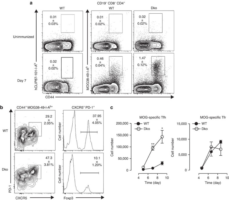
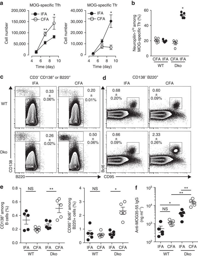
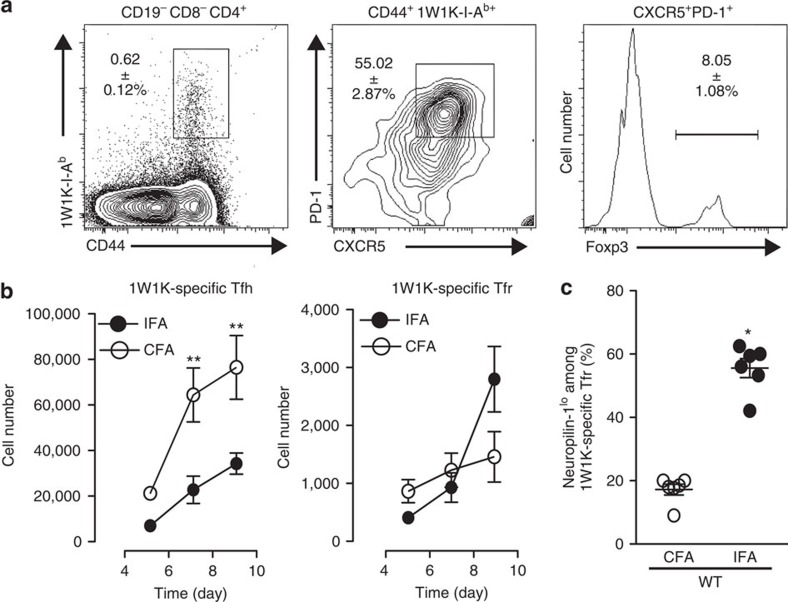
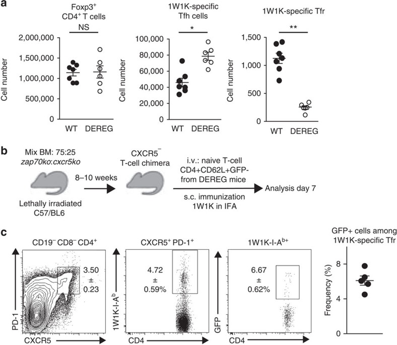
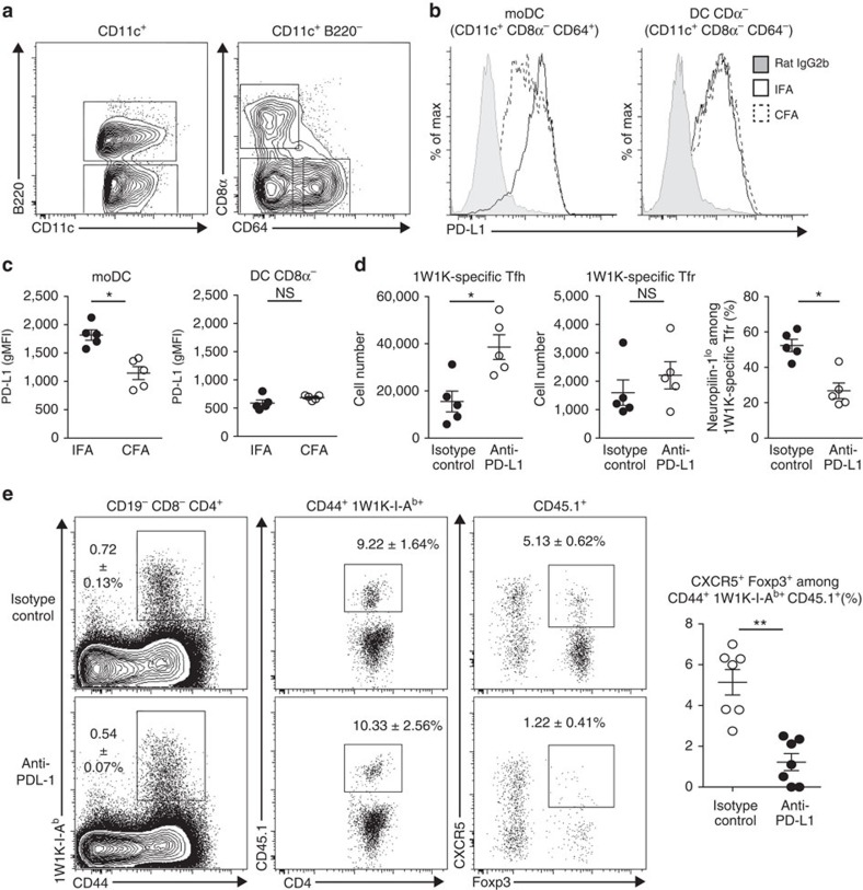
T follicular regulatory (Tfr) cells are a subset of Foxp3(+) regulatory T (Treg) cells that form in response to immunization or infection, which localize to the germinal centre where they control the magnitude of the response. Despite an increased interest in the role of Tfr cells in humoral immunity, many fundamental aspects of their biology remain unknown, including whether they recognize self- or foreign antigen. Here we show that Tfr cells can be specific for the immunizing antigen, irrespective of whether it is a self- or foreign antigen. We show that, in addition to developing from thymic derived Treg cells, Tfr cells can also arise from Foxp3(-) precursors in a PD-L1-dependent manner, if the adjuvant used is one that supports T-cell plasticity. These findings have important implications for Tfr cell biology and for improving vaccine efficacy by formulating vaccines that modify the Tfr:Tfh cell ratio.


Figures

Figure 1
Figure 1. Tfr and Tfh cells are specific for the immunizing Ag.
(a) hCLIP87-101-I-Ab, MOG38-49-I-Ab and Foxp3 staining of CD19CD8αCD4+ dLN (inguinal and periaortic) cells (see Supplementary Fig. 1) from unimmunized WT and Dko mice (top panels) and from WT and Dko mice 7 days after immunization with MOG in CFA (bottom panels). (b) WT and Dko mice were immunized with MOG35-55 emulsified in CFA. dLNs were harvested 7 days after immunization for the detection of MOG-specific activated CD4+ T cells (CD44+ MOG38-49-I-Ab+) and, among them, the follicular cells (CXCR5+ PD-1+). Within the latter, we identified Tfr cells (Foxp3+) in both WT and Dko mice. (c) The mean absolute numbers of total MOG-specific Tfh cells and MOG-specific Tfr cells over time in dLN after immunization of WT (solid circle) or Dko mice (open circle); error bars represent s.e.m. In a,b the numbers in the dot plots represent the mean±s.e.m. Data shown are from a single experiment with five mice per time point and are representative of three independent experiments. Data were analysed using the non-parametric Mann–Whitney test. *P<0.05; **P<0.01.
Figure 2
Figure 2. Immunization with MOG in IFA induces increased numbers of Tfr cells that are Neuropilin-1lo.
Dko mice were immunized with MOG35-55 emulsified in CFA or IFA. (a) Absolute numbers of total MOG-specific Tfh cells and Tfr cells of Dko mice immunized with CFA (open circle, as in Fig. 1c) or IFA (solid circle) at indicated time in dLN. (b) Neuropilin-1lo expression among MOG-specific Tfr cells 7 days after immunization of WT and Dko mice with MOG35-55 emulsified in CFA (white circles) or IFA (black circles). Data shown are from a single experiment (five mice per group) and are representative of three independent experiments. (c) CD138 and B220 staining of CD3 CD138+ or B220+ dLN cells (see Supplementary Fig. 4) from WT and Dko mice 12 days after immunization with MOG in IFA or CFA. (d) CD95 and Bcl6 staining of CD138 B220+ cells. (e) Frequencies of total plasma cells and GC B cells of WT and Dko mice immunized with IFA (solid circle) and CFA (open circle) in dLN 12 days after MOG immunization. (f) Quantity of MOG-specific Ig in the sera of immunized WT and Dko mice estimated using ELISA 12 days after MOG immunization. Data shown are from a single experiment with five mice per group and are representative of two independent experiments. Data were analysed using the non-parametric Mann–Whitney test. ns, not significant, *P<0.05, **P<0.01, mean±s.e.m.
Figure 3
Figure 3. Immunization with a non-self Ag in IFA induces increased numbers of Tfr.
(a) WT mice immunized with 1W1K emulsified in CFA or IFA. dLNs were harvested for the detection of 1W1K-specific CD4+ T cells (CD44+ 1W1K-I-Ab+) and follicular cells (CXCR5+ PD-1+). Tfr cells (1W1K-I-Ab+ CXCR5+ PD-1+ Foxp3+) were identified within total 1W1K-specific follicular cells. (b) Absolute numbers of total 1W1K-specific Tfh cells and Tfr cells over time in dLN after immunization with CFA (open circle) or IFA (solid circle). (c) Neuropilin-1lo expression among 1W1K-specific Tfr cells 7 days after immunization with CFA (white circles) or IFA (black circles). Data are from one experiment of six mice per strain and are representative of three independent experiments. Data were analysed using the non-parametric Mann–Whitney test *P<0.05, **P<0.01, mean±s.e.m.
Figure 4
Figure 4. Naive CD4+ T cells can become Tfr cells.
DEREG and WT mice were immunized with 1W1K emulsified in IFA and were treated with DTx on days 0 and +1. (a) Absolute numbers of total Foxp3+ CD4+ T cells, 1W1K-specific Tfh cells and 1W1K-specific Tfr cells in dLN 7 days after immunization in WT (white circles) and DEREG mice (black circles). Data are representative of six mice per strain. Data were analysed using the non-parametric Mann–Whitney test *P<0.05, **P<0.01, mean±s.e.m. (b) Experimental outline. (c) Flow cytometric dot plots showing follicular T cells (CXCR5+ PD-1+), 1W1K-specific follicular cells (1W1K-IAb+) and 1W1K-specific Tfr cells (GFP+) in the dLN 7 days post immunization with 1W1K in IFA. Data shown are from one experiment with five mice and are representative of two independent experiments.
Figure 5
Figure 5. PD-L1 regulates the formation of induced Tfr cells.
(a) Flow cytometric contour plots showing DC in the dLN 48 h post immunization of WT mice with Ea-OVA in IFA or CFA. (b) Histograms showing PD-L1 expression at the surface of conventional CD8α DC and moDC. (c) Geometric mean fluorescence intensity of PD-L1 expression on the surface of conventional CD8α DC and moDC. Data are from one experiment of five mice per group and are representative of three independent experiments. (d) Number of total 1W1K-specific Tfh and Tfr cells in dLN of WT mice 7 days after immunization with 1W1K emulsified in IFA and treated with anti-PD-L1 (white circles) or isotype control (black circles) on days 0 and +2. Frequency of Neuropilin-1lo among 1W1K-specific Tfr cells. Data are from one experiment of five mice per group and are representative of two independent experiments. (e) Flow cytometric dot plots showing 1W1K-specific CD4+ T cells (CD44+ 1W1K-IAb+), transferred cells among 1W1K-specific CD4+ T cells (CD45.1+) and Tfr cells (CXCR5+ Foxp3+) in the dLN 7 days post immunization with 1W1K in IFA and treated with anti-PD-L1 (white circles) or isotype control (black circles) on days 0 and +2. Data shown are from one experiment with seven mice and are representative of two independent experiments. Data were analysed using the non-parametric Mann–Whitney test. *P<0.05, mean±s.e.m.

References

    1. Fazilleau N., Mark L., McHeyzer-Williams L. J. & McHeyzer-Williams M. G. Follicular helper T cells: lineage and location. Immunity 30, 324–335 (2009). - PMC - PubMed
    1. Chan T. D. & Brink R. Affinity-based selection and the germinal center response. Immunol. Rev. 247, 11–23 (2012). - PubMed
    1. Vinuesa C. G., Sanz I. & Cook M. C. Dysregulation of germinal centres in autoimmune disease. Nat. Rev. Immunol. 9, 845–857 (2009). - PubMed
    1. Vanderleyden I., Linterman M. A. & Smith K. G. Regulatory T cells and control of the germinal centre response. Arthritis Res. Ther. 16, 471 (2014). - PMC - PubMed
    1. Lim H. W., Hillsamer P. & Kim C. H. Regulatory T cells can migrate to follicles upon T cell activation and suppress GC-Th cells and GC-Th cell-driven B cell responses. J. Clin. Invest. 114, 1640–1649 (2004). - PMC - PubMed

Publication types

LinkOut - more resources