. 2016 Feb;49(1):48-57.
doi: 10.1111/cpr.12231. Epub 2016 Jan 27.

Progesterone modulates endothelial progenitor cell (EPC) viability through the CXCL12/CXCR4/PI3K/Akt signalling pathway

Affiliations

Progesterone modulates endothelial progenitor cell (EPC) viability through the CXCL12/CXCR4/PI3K/Akt signalling pathway

Peng Yu et al. Cell Prolif. 2016 Feb.

Abstract

Objectives: Progesterone treatment can effectively increase levels of circulating endothelial progenitor cells (EPCs) and improve neurological functional outcome in a traumatic brain injury (TBI) rat model. However, the mechanisms of progesterone's effects on EPC viability remain elusive. The CXCL12/CXCR4 (CXC chemokine ligand 12/CXC chemokine receptor 4) signalling pathway regulates cell proliferation; we hypothesize that it mediates progesterone-induced EPC viability.

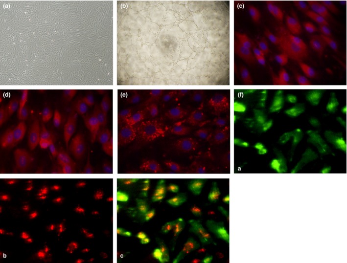
Materials and methods: EPCs were isolated from bone marrow-derived mononuclear cells (BM-MNCs) and treated with progesterone (5, 10 and 100 nm). MTS assay was used to investigate EPC viability. Protein expression was examined by Western blotting, ELISA assay and flow cytometry. Cell membrane and cytoplasm proteins were extracted with membrane and cytoplasm protein extraction kits. CXCR4 antagonist (AMD3100) and phosphatidylinositol 3-kinases (PI3K) antagonist (LY294002) were used to characterize underlying mechanisms.

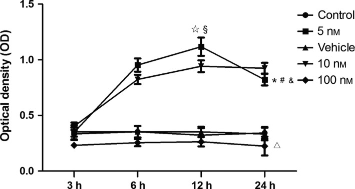
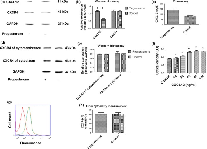
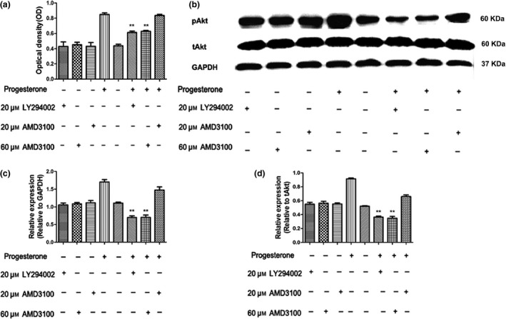
Results: Progesterone-induced EPC viability was time- and dose-dependent. Administration of progesterone facilitated EPC viability and increased expression of CXCL12 and phosphorylated Akt (also known as protein kinase B, pAkt) activity (P < 0.05). Progesterone did not regulate CXCR4 protein expression in cultured EPC membranes or cytoplasm. However, progesterone-induced EPC viability was significantly attenuated by AMD3100 or LY294002. Inhibition of the signalling pathway with AMD3100 and LY294002 subsequently reduced progesterone-induced CXCL12/CXCR4/PI3K/pAkt signalling activity.

Conclusions: The CXCL12/CXCR4/PI3K/pAkt signalling pathway increased progesterone-induced EPC viability.


Figures

Figure 1
Figure 1
(a) Typical morphological appearance of EPC s in culture (10× objective lens). Typical homogeneous and cobblestone‐like morphology of cells after 14 days culture. (b) Tube formation capacity on Matrigel matrix (10 × 4 objective lens). (c–f) Representative immunofluorescence staining of endothelial cell markers including stem cell marker CD34 (c, red), KDR (d, red), vWF (e, red). Cell nuclei were counterstained with DAPI in blue (40x). (f) The cultured cells were able to bind UEA‐1‐FITC (f‐(a), green) and take up DiI‐acLDL (f‐(b), red), (f‐(c), two‐colour overlay)
Figure 2
Figure 2
Concentration‐ and time‐dependent effects of progesterone on EPC viability. Low concentration of progesterone (5 nm) enhanced EPC viability to the greatest extent (*P < 0.05 versus control, vehicle, 10 and 100 nm at 12 h) when incubation was sustained for a short term (no more than 12 h, § P < 0.05 versus 3, 6 and 24 h at 5 nm concentration). High‐dose progesterone treatment (100 nm) dramatically impaired viability of EPCs compared to control. There was no statistical difference in OD values between vehicle and control groups.*Two‐way ANOVA: Significant effect among 3, 6, 12 and 24 h in 5 nm group (P < 0.01) #Two‐way ANOVA: Significant treatment effect between 5 nm group and 10 nm group (P < 0.05), &Two‐way ANOVA: treatment effect between 5 nm group and control (P < 0.05), Two‐way ANOVA: Impaired treatment effect between 100 nm group and control (P < 0.05).
Figure 3
Figure 3
(a) Treating EPC s with progesterone significantly increased levels of CXCL 12 but not CXCR 4 expression. (b) Quantitative analysis of the relative expression of CXCL12 and CXCR4 according to expression of the GAPDH. *P < 0.05 versus control. (c) CXCL12 expression in cultured EPC supernatant.**P < 0.01 versus control. (d) Western blot analysis of CXCR4 protein expressed in cytoplasm and cytomembranes, suggesting that progesterone treatment did not change CXCR4 levels in either. (e) Relative expression of CXCR4 proteinexpressed in cytoplasm and cytomembranes was analysed on the basis of CXCR4/GAPDH ratio. (f) Effect of CXCL12 on EPC viability was measured by MTS. *P < 0.05 versus control, **P < 0.01 versus control. (g) Flow cytometric measurement: progesterone treatment did not increase CXCR4 expression (blue parabola) compared to the non‐treatment group (yellow parabola). Red parabola is the control group without CXCR4 antibody. (H)The percentage of CXCR4+ EPCs in total EPCs after progesterone treatment was measured by flow cytometry.
Figure 4
Figure 4
(a) Progesterone treatment increased EPC viability, while AMD 3100 (60 μ m ) or LY 294002 (20 μ m ) treatment significantly attenuated progesterone‐induced EPC viability. Inhibitors alone had no potential effect on EPC viability. **Two‐way ANOVA: P < 0.01 versus the progesterone group. (b) Western blot analysis indicated that progesterone treatment increased pAkt activity while AMD3100 or LY294002 treatment reduced progesterone‐induced pAkt activity. Treating with inhibitors alone did not change pAkt. (c) Quantitative analysis of the relative expression of pAkt according to the expression of the GAPDH. **Two‐way ANOVA: P < 0.01 versus progesterone group. (d) Relative expression of pAkt was analysed on the basis of pAkt/total Akt ratio. **Two‐way ANOVA: P < 0.01 versus progesterone group.

References

    1. Hu Z, Li Y, Fang M, Wai MS, Yew DT (2009) Exogenous progesterone: a potential therapeutic candidate in CNS injury and neurodegeneration. Curr. Med. Chem. 16, 1418–1425. - PubMed
    1. Peluso JJ, Liu X, Saunders MM, Claffey KP, Phoenix K (2008) Regulation of ovarian cancer cell viability and sensitivity to cisplatin by progesterone receptor membrane component‐1. J. Clin. Endocrinol. Metab. 93, 1592–1599. - PubMed
    1. Peluso JJ, Yuan A, Liu X, Lodde V (2013) Plasminogen activator inhibitor 1 RNA‐binding protein interacts with progesterone receptor membrane component 1 to regulate progesterone's ability to maintain the viability of spontaneously immortalized granulosa cells and rat granulosa cells. Biol. Reprod. 88, 20. - PMC - PubMed
    1. Li Z, Wang B, Kan Z, Zhang B, Yang Z, Chen J et al (2012) Progesterone increases circulating endothelial progenitor cells and induces neural regeneration after traumatic brain injury in aged rats. J. Neurotrauma 29, 343–353. - PMC - PubMed
    1. Xue S, Zhang HT, Zhang P, Luo J, Chen ZZ, Jang XD et al (2010) Functional endothelial progenitor cells derived from adipose tissue show beneficial effect on cell therapy of traumatic brain injury. Neurosci. Lett. 473, 186–191. - PubMed

Publication types

MeSH terms

LinkOut - more resources