. 2016 Jul 7;8(25):12544-52.
doi: 10.1039/c5nr07796f. Epub 2016 Jan 28.

Enhanced performance of macrophage-encapsulated nanoparticle albumin-bound-paclitaxel in hypo-perfused cancer lesions

Affiliations

Enhanced performance of macrophage-encapsulated nanoparticle albumin-bound-paclitaxel in hypo-perfused cancer lesions

Fransisca Leonard et al. Nanoscale. .

Abstract

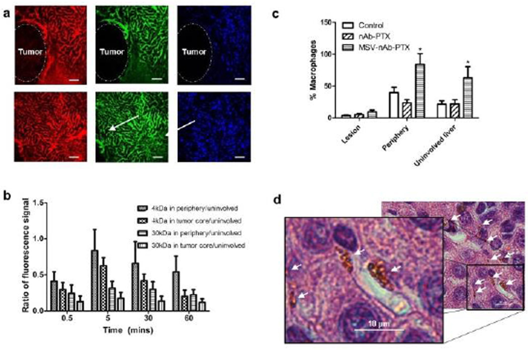
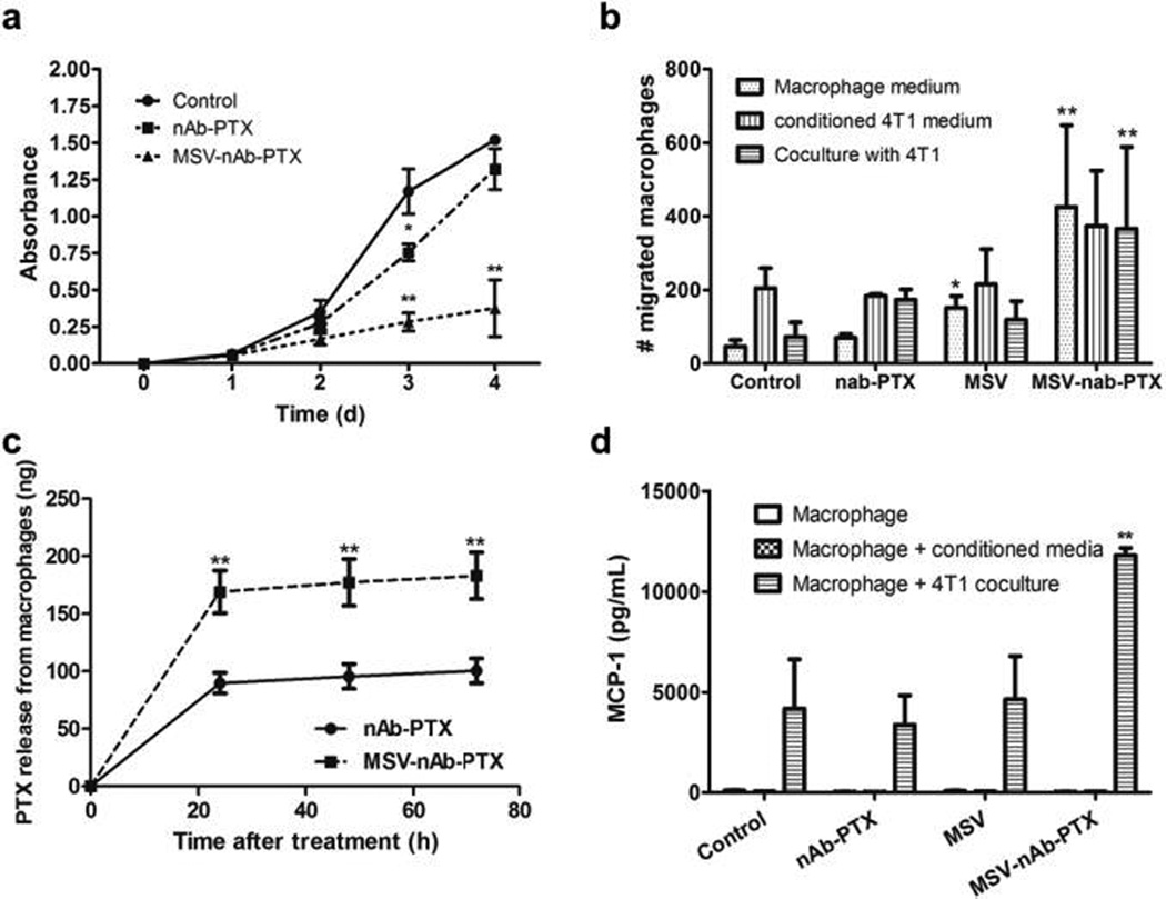
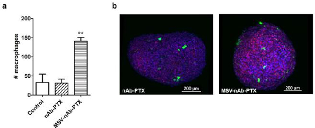
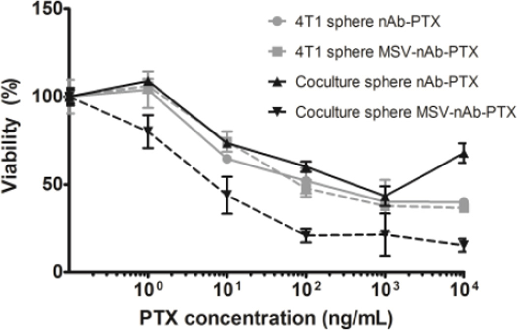
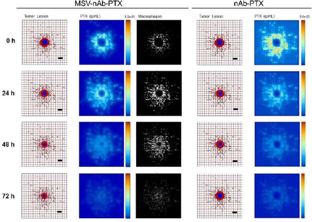
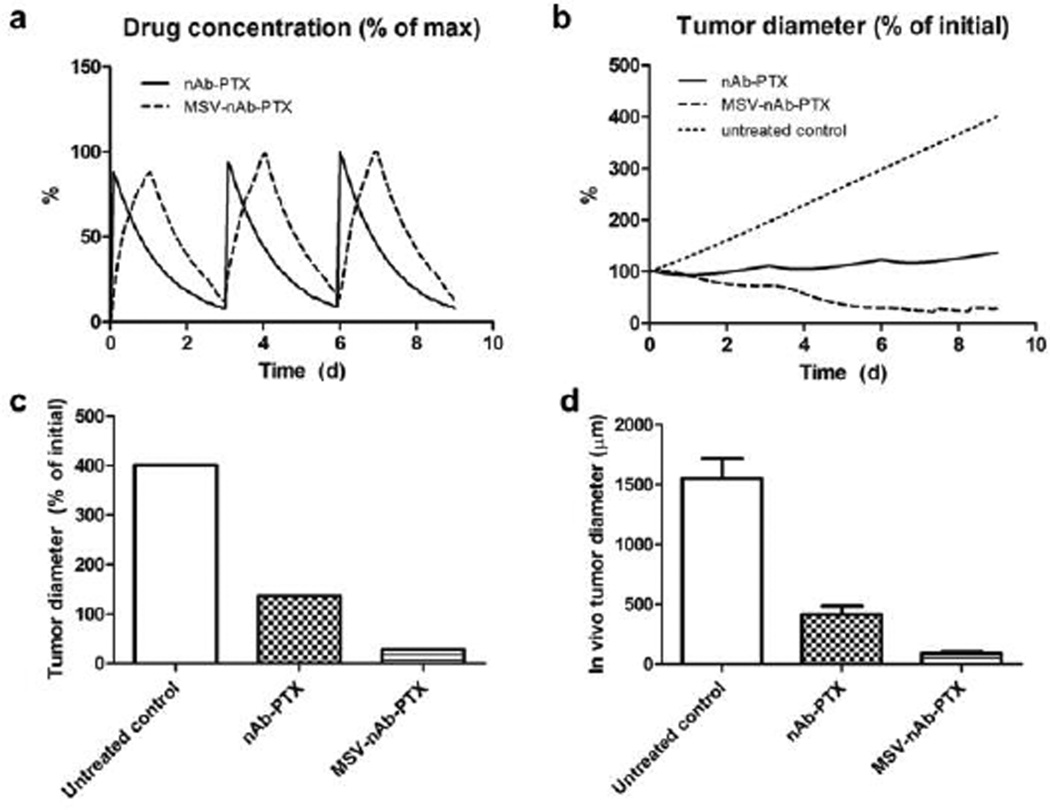
Hypovascularization in tumors such as liver metastases originating from breast and other organs correlates with poor chemotherapeutic response and higher mortality. Poor prognosis is linked to impaired transport of both low- and high-molecular weight drugs into the lesions and to high washout rate. Nanoparticle albumin-bound-paclitaxel (nAb-PTX) has demonstrated benefits in clinical trials when compared to paclitaxel and docetaxel. However, its therapeutic efficacy for breast cancer liver metastasis is disappointing. As macrophages are the most abundant cells in the liver tumor microenvironment, we design a multistage system employing macrophages to deliver drugs into hypovascularized metastatic lesions, and perform in vitro, in vivo, and in silico evaluation. The system encapsulates nAb-PTX into nanoporous biocompatible and biodegradable multistage vectors (MSV), thus promoting nAb-PTX retention in macrophages. We develop a 3D in vitro model to simulate clinically observed hypo-perfused tumor lesions surrounded by macrophages. This model enables evaluation of nAb-PTX and MSV-nab PTX efficacy as a function of transport barriers. Addition of macrophages to this system significantly increases MSV-nAb-PTX efficacy, revealing the role of macrophages in drug transport. In the in vivo model, a significant increase in macrophage number, as compared to unaffected liver, is observed in mice, confirming the in vitro findings. Further, a mathematical model linking drug release and retention from macrophages is implemented to project MSV-nAb-PTX efficacy in a clinical setting. Based on macrophage presence detected via liver tumor imaging and biopsy, the proposed experimental/computational approach could enable prediction of MSV-nab PTX performance to treat metastatic cancer in the liver.


Figures

Figure 1
Figure 1
Characterization of breast cancer liver metastasis in mice: a) Intravital microscopy images of tumor lesion (upper panel) and unaffected liver (lower panel) perfused with 3kDA (red), 40kDA (green) dextrans. Scale bar=100µm. b) Quantification of dextran marker penetration within the periphery (up to 100µm depth into the tumor) or the tumor core normalized to the dextran fluorescence signal in uninvolved liver. c) Quantification of macrophages in vivo based on the location within the lesion, on the periphery of the lesion (the area within 50µm border outside the lesion), or in the uninvolved liver. d) Microscope image of particles taken up by macrophages in the liver (arrows). Scale bar=10µm. n=9, mean±SD, *indicates significant difference (p<0.05) as compared to untreated control.
Figure 2
Figure 2
Studies with 2D transwell co-culture of macrophages (apical side) and breast cancer cells (basolateral side). a) Inhibition of 4T1 breast cancer cells proliferation in co-culture with macrophages pretreated with MSV-nAb-PTX, nAb-PTX and PBS (no treatment) as evaluated by NIS Elements image analysis. b) Migration of macrophages from the apical to basolateral compartment of the migration chamber when the tumor cells were incubated in the basolateral compartment. Macrophages were pretreated with nAb-PTX, MSV or MSV-nAb-PTX for 4h prior to seeding on the apical compartment. Migration was analysed 2d after co-culture. c) Amount of active compound PTX released from the nAb-PTX and MSV-nAb-PTX pre-treated macrophages for 24–72h as assessed by LC/MS-MS. d) Monocyte Chemoattractant Protein-1(MCP-1) released by the cancer cells in the co-culture study in transwell setup. n=6 for a, n=3 for b-d, mean±SD, *indicates significant difference (p<0.05), **very significant difference (p<0.01) to untreated control, except c: to nab=PTX.
Figure 3
Figure 3
Macrophage accumulation and migration into 4T1 tumor spheres: a) Analysis of the total cell numbers associated with the spheroid; b) 3D reconstruction of confocal laser scanning microscopy images of 4T1 spheres (blue) invaded by macrophages (green) and dead cells was imaged by DRAQ7 (red). Macrophages were pre-treated with nAb-PTX or MSV-nAb-PTX.
Figure 4
Figure 4
Effects of nAb-PTX and MSV-nAb-PTX on breast cancer cells (4T1) grown in a monoculture in 3D sphere (grey lines) or in co-culture with primary human macrophages (black lines). Solid lines indicate treatment with nAb-PTX and dotted lines with MSV-nAb-PTX. Data were normalized to control untreated cancer cell 3D spheres and cancer cell sphere-macrophage co-culture. n=6, mean±SD.
Figure 5
Figure 5
Simulation of breast cancer liver metastasis therapy with MSV-nab-PTX (left) and nab-PTX (right): Presentation of tumor lesions, concentration of drug (PTX) released from either macrophages or vessels, and macrophages (only in the case of MSV-nab-PTX). Tumor lesions at the indicated times post treatment initiation. In this simulation, the viable tumor tissue (red) encloses a hypoxic region (blue) without necrosis. The dense capillary network in the liver is modelled by the rectangular grid, with a few irregular sprouts generated through angiogenesis. Size of the lesion is simulated in parallel with drug distribution. PTX represents drug concentration (gm/mL) in the lesion. MSV-nab-PTX are retained in the lesion through the interaction with macrophages while drug is slowly released in the proximity of the tumor cells. Individual macrophages (white) are recruited to vicinity of the lesion based on chemoattraction to hypoxic regions. Bar, 200µm.
Figure 6
Figure 6
Comparison of simulation results (a–c) to response observed in vivo due to repeated therapy over the course of 9d, showing a) simulated drug (as % of maximum blood levels) and b) simulated tumor effect (as % of initial lesion diameter) after nAb-PTX and MSV-nAb-PTX injection. In all cases, therapy is simulated to begin on Days 0, 3, and 6. c) Simulated tumor diameter after 3 treatments as percentage of initial tumor. d) Comparable results from in vivo tumor after 3 treatments as reported in our recent publication. The longer-acting and spatially focused drug release with macrophages achieves a more pronounced regression over the course of therapy than with bolus injection.

References

    1. Selzner M, Morse MA, Vredenburgh JJ, Meyers WC, Clavien PA. Surgery. 2000;127:383–389. - PubMed
    1. Wyld L, Gutteridge E, Pinder SE, James JJ, Chan SY, Cheung KL, Robertson JFR, Evans AJ. Br. J. Cancer. 2003;89:284–290. - PMC - PubMed
    1. van den Eynden GG, Majeed AW, Illemann M, Vermeulen PB, Bird NC, Høyer-Hansen G, Eefsen RL, Reynolds AR, Brodt P. Cancer Res. 2013;73:2031–2043. - PubMed
    1. Stessels F, van den Eynden G, van der Auwera I, Salgado R, van den Heuvel E, Harris AL, Jackson DG, Colpaert CG, van Marck EA, Dirix LY, Vermeulen PB. Br. J. Cancer. 2004;90:1429–1436. - PMC - PubMed
    1. Pezzella F, Pastorino U, Tagliabue E, Andreola S, Sozzi G, Gasparini G, Menard S, Gatter KC, Harris AL, Fox S, Buyse M, Pilotti S, Pierotti M, Rilke F. Am. J. Pathol. 1997;151:1417–1423. - PMC - PubMed

Substances