. 2016 Jan 27;36(4):1336-46.
doi: 10.1523/JNEUROSCI.1171-15.2016.

Platelet-Activating Factor Receptors Mediate Excitatory Postsynaptic Hippocampal Injury in Experimental Autoimmune Encephalomyelitis

Affiliations

Platelet-Activating Factor Receptors Mediate Excitatory Postsynaptic Hippocampal Injury in Experimental Autoimmune Encephalomyelitis

Matthew J Bellizzi et al. J Neurosci. .

Abstract

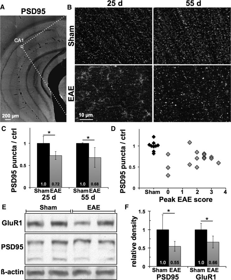
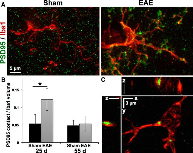
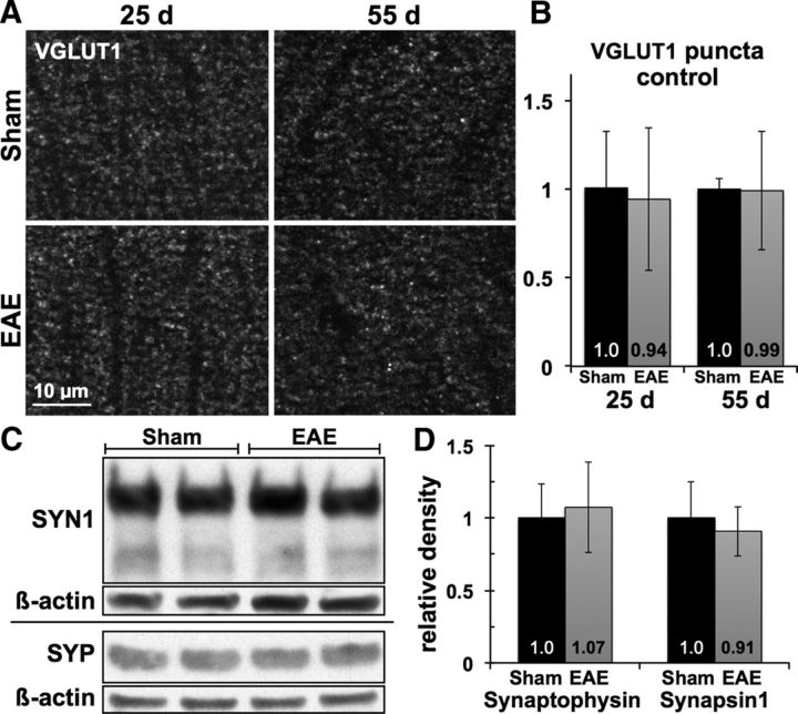
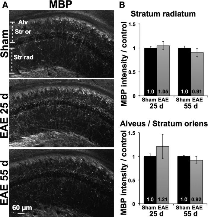
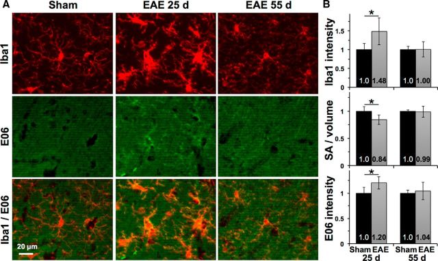
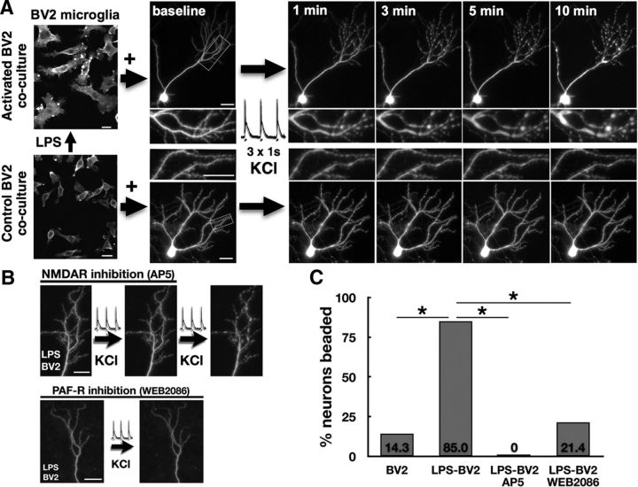
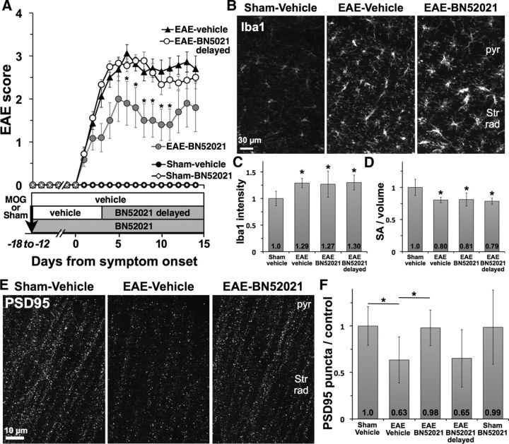
Gray matter degeneration contributes to progressive disability in multiple sclerosis (MS) and can occur out of proportion to measures of white matter disease. Although white matter pathology, including demyelination and axon injury, can lead to secondary gray matter changes, we hypothesized that neurons can undergo direct excitatory injury within the gray matter independent of these. We tested this using a model of experimental autoimmune encephalomyelitis (EAE) with hippocampal degeneration in C57BL/6 mice, in which immunofluorescent staining showed a 28% loss of PSD95-positive excitatory postsynaptic puncta in hippocampal area CA1 compared with sham-immunized controls, despite preservation of myelin and VGLUT1-positive excitatory axon terminals. Loss of postsynaptic structures was accompanied by appearance of PSD95-positive debris that colocalized with the processes of activated microglia at 25 d after immunization, and clearance of debris was followed by persistently reduced synaptic density at 55 d. In vitro, addition of activated BV2 microglial cells to hippocampal cultures increased neuronal vulnerability to excitotoxic dendritic damage following a burst of synaptic activity in a manner dependent on platelet-activating factor receptor (PAFR) signaling. In vivo treatment with PAFR antagonist BN52021 prevented PSD95-positive synapse loss in hippocampi of mice with EAE but did not affect development of EAE or local microglial activation. These results demonstrate that postsynaptic structures can be a primary target of injury within the gray matter in autoimmune neuroinflammatory disease, and suggest that this may occur via PAFR-mediated modulation of activity-dependent synaptic physiology downstream of microglial activation.

Significance statement: Unraveling gray matter degeneration is critical for developing treatments for progressive disability and cognitive impairment in multiple sclerosis (MS). In a mouse model of MS, we show that neurons can undergo injury at their synaptic connections within the gray matter, independent of the white matter pathology, demyelination, and axon injury that have been the focus of most current and emerging treatments. Damage to excitatory synapses in the hippocampus occurs in association with activated microglia, which can promote excitotoxic injury via activation of receptors for platelet-activating factor, a proinflammatory signaling molecule elevated in the brain in MS. Platelet-activating factor receptor blockade protected synapses in the mouse model, identifying a potential target for neuroprotective treatments in MS.

Keywords: excitotoxicity; microglia; multiple sclerosis; neurodegeneration; neuroprotection; platelet-activating factor.


Figures

Figure 1.
Figure 1.
Loss of excitatory postsynaptic structures in EAE hippocampus occurs independent of motor deficits. A, Hippocampal sections immunostained for excitatory postsynaptic marker PSD95 were imaged within the stratum radiatum of area CA1 (box). B, High-power images show that punctate staining for PSD95 was disrupted in EAE mice, with appearance of PSD95-positive material in a nonpunctate pattern at 25 dpi that was cleared by 55 dpi. C, Density of PSD95-positive puncta was decreased by 28 ± 9% and 32 ± 22% in EAE mice at 25 and 55 d compared with age- and sex-matched sham-immunized controls. D, Loss of hippocampal PSD95 puncta at either time point did not correlate with peak severity of motor deficits in EAE mice (Spearman's ρ = 0.16, p = 0.56). E, Western blots showed decreases in excitatory postsynaptic proteins PSD95 and GluR1 in hippocampal homogenates from EAE versus sham mice at 23 dpi. All protein bands are from the same gel. F, PSD95 levels normalized to β-actin were decreased by 45 ± 13% in EAE hippocampi compared with sham controls, and GluR1 levels decreased by 34 ± 16%. *p < 0.05.
Figure 2.
Figure 2.
Microglia contact and engulf excitatory postsynaptic structures in EAE hippocampus. A, Loss of PSD95-positive puncta in EAE mice at 25 d coincided with accumulation of PSD95-positive material along the hypertrophic processes of Iba1-positive microglia. B, Volume of PSD95-positive structures in contact with microglia was increased >2-fold in EAE hippocampi at 25 d and returned to levels similar to sham-immunized controls by 55 d. *p = 0.004. C, PSD95-positive inclusions can be seen within Iba1-positive microglia in EAE hippocampus at day 55.
Figure 3.
Figure 3.
Excitatory axon terminals remain intact in EAE hippocampus. A, B, Punctate immunostaining for excitatory presynaptic marker VGLUT1 remained undisturbed within the CA1 stratum radiatum of EAE mice compared with sham-immunized controls, with no change in density or staining pattern at 25 or 55 dpi. C, D, Levels of presynaptic proteins synapsin-1 and synaptophysin were similar in hippocampal homogenates from EAE versus sham-immunized mice at 23 dpi. Horizontal divider indicates separate gels.
Figure 4.
Figure 4.
Hippocampal myelin is preserved in EAE. A, Immunostaining for myelin basic protein (MBP) was preserved in the CA1 stratum radiatum (Str rad) as well as along pyramidal neuron axons in the stratum oriens (Str or) and alveus (Alv) in EAE mice compared with sham controls (25 dpi sham pictured). B, MBP intensity was not decreased in either region at 25 dpi, and any attenuation at 55 dpi was mild and not statistically significant.
Figure 5.
Figure 5.
Microglial activation and oxidation in EAE hippocampus. A, Iba1-positive microglia developed a hypertrophic morphology in EAE mice at 25 dpi and an intermediate morphology with shorter and less-ramified processes at 55 dpi compared with those from sham-immunized controls. Top, The 25 dpi sham. E06 immunostaining for oxidized phospholipids was increased within and around microglia in EAE at 25 dpi and was attenuated at 55 dpi with residual staining in some microglia. Bottom, Thresholding applied equally to E06 images. B, Segmentation analysis shows increased Iba1 intensity, lower surface area-to-volume ratio, and increased E06 intensity within microglia in EAE at 25 dpi. *p < 0.025 versus sham.
Figure 6.
Figure 6.
Activated BV2 microglia promote activity-dependent postsynaptic injury. A, GFP-expressing neurons in hippocampal cultures maintained normal dendrite morphology 24 h after addition of BV2 microglial cells activated by LPS (500 ng/ml for 6 h) or unstimulated controls. A burst of synaptic activity triggered by pulses of depolarizing KCl (90 mm, three 1 s pulses every 10 s) caused progressive and sustained dendritic beading in neurons cocultured with activated BV2s but not controls (boxed areas enlarged in insets). B, KCl-triggered dendritic beading is reversibly blocked by NMDA receptor antagonist AP5 (10 μm) and is prevented by addition of PAFR antagonist WEB2086 (10 μm) to neurons cocultured with LPS-activated BV2s. C, Rates of KCl-triggered dendritic beading, increased in neurons cocultured with LPS-activated BV2s, were abolished by AP5 and were reduced to control levels by WEB2086. *p < 0.001. Scale bars, 20 μm.
Figure 7.
Figure 7.
PAFR antagonism protects hippocampal synapses in EAE. A, Treatment with PAFR antagonist BN52021 beginning the day after immunization (EAE-BN52021) did not prevent development of EAE but modestly reduced severity of motor deficits compared with EAE mice treated with vehicle or BN52021 delayed until after onset of motor deficits. *p < 0.05 versus EAE vehicle. B, BN52021 treatment did not affect activation of Iba1-positive microglia in CA1 hippocampus. pyr, Pyramidal cell layer; Str Rad, stratum radiatum. C, D, Microglial Iba1 intensity was increased and surface area-to-volume ratio was decreased in all groups of EAE mice, whether treated with vehicle, BN52021 from day 1, or BN52021 delayed. *p < 0.05 versus sham-vehicle. E, F, PSD95-positive synaptic puncta were decreased by 37% in hippocampi of vehicle-treated EAE mice, and maintained at control levels in EAE mice treated with BN52021. Delayed BN52021 treatment did not protect PSD95-positive synapses in EAE, and BN52021 treatment had no effect on puncta in sham-immunized controls. *p < 0.05 versus sham-vehicle.

References

    1. Amato MP, Bartolozzi ML, Zipoli V, Portaccio E, Mortilla M, Guidi L, Siracusa G, Sorbi S, Federico A, De Stefano N. Neocortical volume decrease in relapsing-remitting MS patients with mild cognitive impairment. Neurology. 2004;63:89–93. doi: 10.1212/01.WNL.0000129544.79539.D5. - DOI - PubMed
    1. Bellizzi MJ, Lu SM, Masliah E, Gelbard HA. Synaptic activity becomes excitotoxic in neurons exposed to elevated levels of platelet-activating factor. J Clin Invest. 2005;115:3185–3192. doi: 10.1172/JCI25444. - DOI - PMC - PubMed
    1. Brown GC, Neher JJ. Microglial phagocytosis of live neurons. Nat Rev Neurosci. 2014;15:209–216. doi: 10.1038/nrn3710. - DOI - PubMed
    1. Callea L, Arese M, Orlandini A, Bargnani C, Priori A, Bussolino F. Platelet activating factor is elevated in cerebral spinal fluid and plasma of patients with relapsing-remitting multiple sclerosis. J Neuroimmunol. 1999;94:212–221. doi: 10.1016/S0165-5728(98)00246-X. - DOI - PubMed
    1. Centonze D, Muzio L, Rossi S, Cavasinni F, De Chiara V, Bergami A, Musella A, D'Amelio M, Cavallucci V, Martorana A, Bergamaschi A, Cencioni MT, Diamantini A, Butti E, Comi G, Bernardi G, Cecconi F, Battistini L, Furlan R, Martino G. Inflammation triggers synaptic alteration and degeneration in experimental autoimmune encephalomyelitis. J Neurosci. 2009;29:3442–3452. doi: 10.1523/JNEUROSCI.5804-08.2009. - DOI - PMC - PubMed

Publication types

MeSH terms

Substances

LinkOut - more resources