. 2016 Jan 27;36(4):1410-5.
doi: 10.1523/JNEUROSCI.2456-15.2016.

Neurodegeneration Triggers Peripheral Immune Cell Recruitment into the Forebrain

Affiliations

Neurodegeneration Triggers Peripheral Immune Cell Recruitment into the Forebrain

Miriam Scheld et al. J Neurosci. .

Abstract

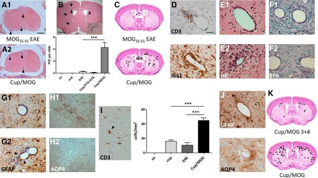
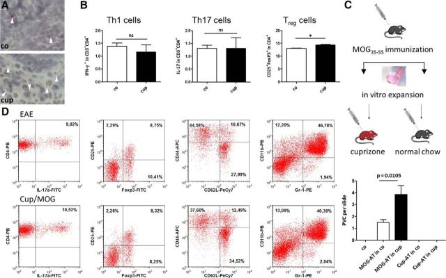
Brain-intrinsic degenerative cascades have been proposed to be an initial factor driving lesion formation in multiple sclerosis (MS). Here, we identify neurodegeneration as a potent trigger for peripheral immune cell recruitment into the mouse forebrain. Female C57BL/6 mice were fed cuprizone for 3 weeks, followed by a period of 2 weeks on normal chow to induce the formation of lesion foci in the forebrain. Subsequent immunization with myelin oligodendrocyte glycoprotein 35-55 peptide, which induces myelin autoreactive T cells in the periphery, resulted in massive immune cell recruitment into the affected forebrain. Additional adoptive transfer experiments together with flow cytometry analysis underline the importance of brain-derived signals for immune cell recruitment. This study clearly illustrates the significance of brain-intrinsic degenerative cascades for immune cell recruitment and MS lesion formation. Additional studies have to address the signaling cascades and mechanistic processes that form the top-down communication between the affected brain area, neurovascular unit, and peripheral immune cells.

Significance statement: We identify neurodegeneration as a potent trigger for peripheral immune cell recruitment into the forebrain. Thus, immune cell recruitment might be a second step during the formation of new inflammatory lesions in multiple sclerosis. A better understanding of factors regulating neurodegeneration-induced immune cell recruitment will pave the way for the development of novel therapeutic treatment strategies.

Keywords: cytodegeneration; invasion; trigger.


Figures

Figure 1.
Figure 1.
A, Shows representative H&E-stained sections of the spinal cord (at the peak of the disease, ∼2 weeks after immunization). Arrowheads highlight inflammatory foci. B and C show the number and spatial distribution of perivascular infiltrates in the different treatment groups (H&E staining). D, Composition of perivascular infiltrates. Both CD3+ lymphocytes and Iba1+ monocytes can be found. E shows two distinct lesion types: E1, perivascular lesions in which immune cells are trapped in the enlarged perivascular space; E2, where immune cells invaded the neuropil. Immune cell neuropil invasion induces demyelination [Luxol fast blue (LFB)/periodic acid Schiff; F1, F2], the breakdown of the glia limitans perivascularis (anti-Glial fibrillary acidic protein; G1, G2, see arrows), and astrocyte polarity loss (anti-AQP4; H1, H2). I, Quantification of CD3+ cells into the neuropil. Arrow points at a CD3+ lymphocyte within the neuropil. J shows the integrity of the glia limitans perivascularis 6 weeks after immunization. Anti-Glial fibrillary acidic protein presents as a continuous layer of astrocyte end feet around vessels; focal loss of anti AQP-4 stain is not evident anymore. K shows the cumulative number and spatial distribution of perivascular infiltrates after delayed MOG immunization (i.e., 4 instead of 2 weeks after Cup-induced neurodegeneration).
Figure 2.
Figure 2.
A, Representative ORO-stained cervical lymph nodes. White arrowheads highlight lipid-laden macrophages. B shows relative numbers of Th1, Th17, and Treg cells in cervical lymph nodes before MOG immunization. Note that the immune cell repertoire is not altered as a result of the Cup intoxication. C shows the number of forebrain perivascular infiltrates in different treatment groups. Note that perivascular infiltrates were virtually absent in co animals (i) and in animals in which cultured and stimulated splenocytes, isolated from Cup-exposed animals, were transferred into either co animals (Cup-AT in co) or Cup-intoxicated animals (Cup-AT in cup; ii). In contrast, numerous perivascular infiltrates were present in classical AT experiments. Most importantly, the number of forebrain perivascular infiltrates is greater in animals with Cup-induced neurodegeneration before T-cell transfer (MOG-AT in co vs MOG-AT in cup). PVC, Perivascular cuffs. D, Immune cell repertoire in the spleen, determined by FACS analysis. The following marker sets were used: Th17 cells (IL17+/CD3+/CD4+), Treg cells (CD25+/FoxP3+/CD4+), memory T cells (Tmem; CD62Llow/CD44high/CD4+), and myeloid-derived suppressor cells (CD11b+/Gr1+).

References

    1. Aboul-Enein F, Rauschka H, Kornek B, Stadelmann C, Stefferl A, Brück W, Lucchinetti C, Schmidbauer M, Jellinger K, Lassmann H. Preferential loss of myelin-associated glycoprotein reflects hypoxia-like white matter damage in stroke and inflammatory brain diseases. J Neuropathol Exp Neurol. 2003;62:25–33. - PubMed
    1. Almolda B, González B, Castellano B. Activated microglial cells acquire an immature dendritic cell phenotype and may terminate the immune response in an acute model of EAE. J Neuroimmunol. 2010;223:39–54. doi: 10.1016/j.jneuroim.2010.03.021. - DOI - PubMed
    1. Barnett MH, Prineas JW. Relapsing and remitting multiple sclerosis: pathology of the newly forming lesion. Ann Neurol. 2004;55:458–468. doi: 10.1002/ana.20016. - DOI - PubMed
    1. Baxi EG, DeBruin J, Tosi DM, Grishkan IV, Smith MD, Kirby LA, Strasburger HJ, Fairchild AN, Calabresi PA, Gocke AR. Transfer of myelin-reactive th17 cells impairs endogenous remyelination in the central nervous system of cuprizone-fed mice. J Neurosci. 2015;35:8626–8639. doi: 10.1523/JNEUROSCI.3817-14.2015. - DOI - PMC - PubMed
    1. Clarner T, Diederichs F, Berger K, Denecke B, Gan L, van der Valk P, Beyer C, Amor S, Kipp M. Myelin debris regulates inflammatory responses in an experimental demyelination animal model and multiple sclerosis lesions. Glia. 2012;60:1468–1480. doi: 10.1002/glia.22367. - DOI - PubMed

Publication types

MeSH terms

LinkOut - more resources