. 2016 Jan 28:6:20040.
doi: 10.1038/srep20040.

Xuefu Zhuyu decoction, a traditional Chinese medicine, provides neuroprotection in a rat model of traumatic brain injury via an anti-inflammatory pathway

Affiliations

Xuefu Zhuyu decoction, a traditional Chinese medicine, provides neuroprotection in a rat model of traumatic brain injury via an anti-inflammatory pathway

Zhihua Xing et al. Sci Rep. .

Abstract

Neuroinflammation is central to the pathology of traumatic brain injury (TBI). Xuefu Zhuyu decoction (XFZY) is an effective traditional Chinese medicine to treat TBI. To elucidate its potential molecular mechanism, this study aimed to demonstrate that XFZY functions as an anti-inflammatory agent by inhibiting the PI3K-AKT-mTOR pathway. Sprague-Dawley rats were exposed to controlled cortical impact to produce a neuroinflammatory response. The treatment groups received XFZY (9 g/kg and 18 g/kg), Vehicle group and Sham group were gavaged with equal volumes of saline. The modified neurologic severity score (mNSS) and the Morris water maze test were used to assess neurological deficits. Arachidonic acid (AA) levels in brain tissue were measured using tandem gas chromatography-mass spectrometry. TNF-α and IL-1β levels in injured ipsilateral brain tissue were detected by ELISA. AKT and mTOR expression were measured by western blot analysis. The results indicated that XFZY significantly enhanced spatial memory acquisition. XFZY (especially at a dose of 9 g/kg) markedly reduced the mNSS and levels of AA, TNF-α and IL-1β. Significant downregulation of AKT/mTOR/p70S6K proteins in brain tissues was observed after the administration of XFZY (especially at a dose of 9 g/kg). XFZY may be a promising therapeutic strategy for reducing inflammation in TBI.


Figures

Figure 1
Figure 1. Flow diagram of the experiment.
The experiment was performed on six groups: Vehicle, Sham, 9 g/kg XFZY, 18 g/kg XFZY, Sham 9 g/kg XFZY and Sham 18 g/kg XFZY. The dosage regimen for each group was applied continuously once per day after TBI. The quality control of XFZY was performed using LCMS-IT-TOF. On the 1st, 3rd, 7th and 14th day post-TBI, 8 rats were randomly drawn from each group, except for the sham 9 g/kg XFZY and sham 18 g/kg XFZY groups. The rodents were then sacrificed for sample collection after the neurological function tests. Brain samples were used for biochemical assays and determination of the arachidonic acid by GC-MS. The remaining 12 rats in each above-mentioned group were examined to evaluate the neurological function scores on the 1st, 3rd, 7th, 14th and 21st days after TBI. These 12 rats in each group and 8 rats in each of the sham XFZY groups (9 g/kg or 18 g/kg) underwent MWM testing from the 17th to the 21st day.
Figure 2
Figure 2. Liquid chromatography coupled to high-resolution ion trap and time-of-flight mass spectrometry (LCMS-IT-TOF) analysis for HSYA and Amygdalin determination, which originated from the monarch drug of XFZY.
(A) Prepared Chinese herbal medicines of XFZY in small amounts that are ready for decoction. (B) LCMS-IT-TOF TIC chromatograms of XFZY in positive ESI mode and chromatographic profiles of Amygdalin and HSYA. (C,D) MS and MS of Amygdalin were m/z 458.16 and 296.11, respectively. (E, F) MS and MS of HSYA were m/z 613.17 and 451.12, respectively.
Figure 3
Figure 3. Modified neurologic severity score (mNSS) after TBI or sham injury or TBI with XFZY.
(A) Neurological function was evaluated by mNSS after 1 hour to assess the initial disability and on the 3rd, 7th, 14th, and 21st days after TBI. Treatment with 9 g/kg XFZY significantly lowered the mNSS on the 3rd, 7th, 14th, and 21st days compared with the Vehicle group. Treatment with 18 g/kg XFZY resulted in a significant decrease on the 3rd and 21st days (n = 8/group, data are analyzed by two-way ANOVA and presented as the mean ± SEM. p < 0.05 vs. the Vehicle group). (B) Changes in mNSS (ΔmNSS) were assessed at various time intervals between the 1st day and multiple predetermined time points thereafter, considering that the performance of mNSS was not significant on the 1st day (mean mNSS values: 9 g/kg XFZY group = 12.8 ± 0.4, 18 g/kg XFZY group = 13.2 ± 0.4, Vehicle group = 13.5 ± 0.5, all p > 0.05 compared with each other). Treatment with 9 g/kg XFZY significantly enhanced ΔmNSS over time intervals of 1–3, 1–7, 1–14 and 1–21 days. Treatment with 18 g/kg XFZY significantly increased ΔmNSS during the interval of 1–3 and 1–21 days. The ΔmNSS in the 9 g/kg XFZY group increased significantly during the time intervals of 1–7, 1–14, and 1–21 days compared with the 18 g/kg XFZY group (n = 12/group, data were analyzed with RM ANOVA and are presented as the mean ± SEM. *p < 0.05, #p < 0.01 vs. the Vehicle group. p < 0.05, p < 0.01 vs. 9 g/kg XFZY group).
Figure 4
Figure 4. Effect of XFZY on cognitive outcomes after sham injury or controlled cortical impact (CCI).
(A,B) Sham-injured rats (n = 8/group) were gavaged with 9 g/kg XFZY, 18 g/kg XFZY, or an equivalent amount of saline each day. The Morris water maze (MWM) test was then performed from the 17th to the 21st day after the sham injury. Performance in the MWM revealed no differences between the hidden and visible platform (p > 0.05 for group, repeated-measures ANOVA (RM ANOVA)) or in the probe trials (p > 0.05, B), despite learning the task (p < 0.01 for time, hidden and visible platform trials). (C, D) In the CCI groups (n = 12/group), significant group effects were observed in the hidden platform tests to assess the impact of XFZY treatment (p < 0.01 for the group) with learning the hidden platform paradigms (p < 0.01 for time in each group). The rats that were treated with 9 g/kg XFZY performed significantly better than the vehicle-treated mice in the hidden platform (p < 0.05) and probe trials (p < 0.05). Although there were no significant differences in the hidden platform test, the 18 g/kg XFZY group displayed significantly improved performance in the probe trials compared with the Vehicle group (p < 0.05). No differences in visible platform performance were noted among the CCI groups (p > 0.05 for group). *p < 0.05 versus the Vehicle group.
Figure 5
Figure 5. Quantitative determination of Arachidonic acid (AA) in ipsilateral brain tissues on the 1st, 3rd, 7th, 14th and 21st days after TBI or sham injury or TBI gavaged with oral XFZY.
(A) GC-MS m/z of AA after MeOx-TMS derivatization is 67, 73, 75, 79, 91 and 117. The top diagram shows the mass spectrum of AA in a brain sample, and the bottom diagram shows the mass spectrum of standard AA. (B) GC-MS TICs of AA after MeOx-TMS derivatization. (C) Brain tissue concentration of AA on the 1st, 3rd, 7th, 14th and 21st days after injury. The contents of AA were significantly increased in the Vehicle group compared with the sham group from the 1st to the 14th days post-injury, whereas treatment with 9 g/kg XFZY significantly reduced the increased AA levels compared with the Vehicle group on the 1st, 3rd, 7th, and 14th days. Treatment with 18 g/kg XFZY significantly reduced the increased levels of AA compared with the Vehicle group on the 1st, 3rd, and 7th days. On the 3rd day, treatment with 9 g/kg XFZY significantly reduced the levels of AA in ipsilateral brain tissue compared with 18 g/kg XFZY group (n = 8/group). All of the data were analyzed by two-way ANOVA and are presented as the mean ± SEM. *p < 0.05, #p < 0.01 vs. the Vehicle group. Δp < 0.05 vs. the 9 g/kg XFZY group.
Figure 6
Figure 6. Assay of pro-inflammatory cytokine levels in brain tissue.
(A) The levels of TNF-α in brain lysates from each group. The TNF-α levels were significantly increased in the Vehicle group compared with the Sham group from the 1st to the 14th day post-injury, whereas treatment with 9 g/kg XFZY significantly reduced the increase in TNF-α compared with the Vehicle group on the 1st, 3rd, 7th and 14th days. Treatment with 18 g/kg XFZY significantly reduced the increase in TNF-α levels compared with the Vehicle group on the 1st, 3rd and 7th days (n = 8/group). On the 7th and 14th days, the levels of TNF-α in the 9 g/kg XFZY group were significantly lower than those in the 18 g/kg XFZY group. (B) The levels of IL-1β in brain lysates in each group. The contents of IL-1β were significantly increased in the Vehicle group compared with the Sham group from the 1st to the 14th day post-injury. Treatment with 9 g/kg XFZY significantly reduced the increased levels of IL-1β compared with the Vehicle group on the 1st, 3rd, 7th, and 14th days. Treatment with 18 g/kg XFZY significantly reduced the increased levels of IL-1β compared with the Vehicle group on the 1st, 3rd, 7th, and 14th days. On the 3rd and 7th days, treatment with 9 g/kg XFZY significantly reduced the levels of IL-1β in the ipsilateral brain tissue compared with treatment with 18 g/kg XFZY (n = 8/group). All of the data were analyzed by two-way ANOVA and are presented as the mean ± SEM. *p < 0.05, #p < 0.01 vs. the Vehicle group. p < 0.05 vs. the 9 g/kg XFZY group.
Figure 7
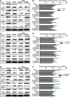
Figure 7. The effects of XFZY on the PI3K-AKT-mTOR signaling pathway after TBI.
(A,B) The levels of p-AKT decreased significantly on the 1st and 3rd days in ipsilateral brain tissue after TBI. Treatment with 9 g/kg XFZY significantly reduced the increase in p-AKT expression from the 1st to the 14th day compared with the Vehicle group. The same trend was observed in the 18 g/kg XFZY group on the 1st, 3rd and 14th days. The expression levels of p-AKT were significantly lower in the 9 g/kg XFZY group compared with the 18 g/kg XFZY group on the 7th and 14th days. No significant changes were detected in total AKT expression between the Sham and Vehicle group. (C,D) p-mTOR was significantly increased from the 1st to the 14th day after TBI. Treatment with 9 g/kg XFZY significantly decreased p-mTOR expression compared with the Vehicle group from the 1st to the 14th day. Treatment with 18 g/kg XFZY significantly reduced p-mTOR on the 1st and 3rd days. mTOR expression in the 9 g/kg XFZY group was significantly reduced compared with the 18 g/kg XFZY group on the 1st, 7th and 14th days. There were no significant changes in total mTOR in comparisons between each group. (E,F) p-p70S6K increased significantly from the 1st to the 14th day after TBI. Treatment with 9 g/kg XFZY significantly reduced p-p70S6K expression on the above days. The same tendency was observed in the 18 g/kg XFZY group on the 1st day. p-p70S6K expression was significantly reduced in the 9 g/kg XFZY group compared with the 18 g/kg XFZY group on the 3rd, 7th and 14th days. No significant changes were detected in total p70S6K expression between the Sham and Vehicle group. Data are the mean ratio between targeting proteins and β-actin and are presented as the percentage of XFZY (or Vehicle)-treated brains over the control Sham-treated brains. The data represent the mean fold induction ± SEM, as analyzed by ANOVA. *p < 0.05, #p < 0.01 vs. the Vehicle group. p < 0.05, p < 0.01 vs. the 9 g/kg XFZY group.
Figure 8
Figure 8. Inflammatory response in the brain after TBI and the pathomechanism of the anti-inflammatory effect of XFZY.
Following TBI, the mechanical injury-stimulated cell membrane releases arachidonic acid (AA), which is metabolized into prostaglandin E2 (PGE2) and prostacyclin (PGI2) by cyclooxygenase-2 (COX-2) and into leukotrienes (LTB4) by 5-lipoxygenase (5-LO). The three inflammatory mediators initiate acute inflammation, including changes in blood flow, increased capillary permeability and inflammatory cell recruitment in the brain injury ambitus zone, including polymorphonuclear leukocytes (i.e., neutrophils) and monocytes. Excess prostaglandins and leukotrienes contribute to chronic inflammation. The neutrophils are activated to further release chemotactic factors, devour necrotic tissue and sterilize bacteria. The influx of monocytes and resident microglial cells develop into macrophages, which secrete pro-inflammatory factors (e.g., TNF-α and IL-1β) and consume foreign bodies, necrotic tissue or apoptotic cells (e.g., efferocytosis). XFZY significantly suppressed the increased levels of blood AA, TNF-α and IL-1β in brain tissue, indicating that XFZY possesses anti-inflammatory effects. To target the PI3K-AKT-mTOR signaling pathway, XFZY significantly reversed the elevated phosphorylation of AKT/mTOR in brain tissue post-TBI, as well as the downstream p70S6K, resulting in a reduced translation ratio of inflammatory factors and exerting anti-inflammatory effects.

References

    1. Feigin V. L. et al.. Incidence of traumatic brain injury in New Zealand: a population-based study. Lancet Neuro. 12, 53–64 (2013). - PubMed
    1. Wright D. W. et al.. Very early administration of progesterone for acute traumatic brain injury. N. Engl. J. Med. 371, 2457–2466 (2014). - PMC - PubMed
    1. Lozano D. et al.. Neuroinflammatory responses to traumatic brain injury: etiology, clinical consequences, and therapeutic opportunities. Neuropsychiatr. Dis. Treat. 11, 97–106 (2015). - PMC - PubMed
    1. Kou K., Hou X. Y., Sun J. D. & Chu K. Current pre-hospital traumatic brain injury management in China. World J. Emerg. Med. 5, 245–254 (2014). - PMC - PubMed
    1. Alluri H. et al.. Blood-brain barrier dysfunction following traumatic brain injury. Metab. Brain. Dis. 30, 1093–1104 (2015). - PubMed

Publication types

MeSH terms

LinkOut - more resources