. 2016 Jan 27:17:29.
doi: 10.1186/s12863-016-0338-9.

Focal exposure of limited lung volumes to high-dose irradiation down-regulated organ development-related functions and up-regulated the immune response in mouse pulmonary tissues

Affiliations

Focal exposure of limited lung volumes to high-dose irradiation down-regulated organ development-related functions and up-regulated the immune response in mouse pulmonary tissues

Bu-Yeo Kim et al. BMC Genet. .

Abstract

Background: Despite the emergence of stereotactic body radiotherapy (SBRT) for treatment of medically inoperable early-stage non-small-cell lung cancer patients, the molecular effects of focal exposure of limited lung volumes to high-dose radiation have not been fully characterized. This study was designed to identify molecular changes induced by focal high-dose irradiation using a mouse model of SBRT.

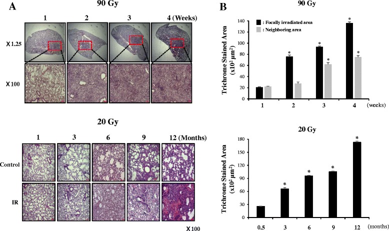
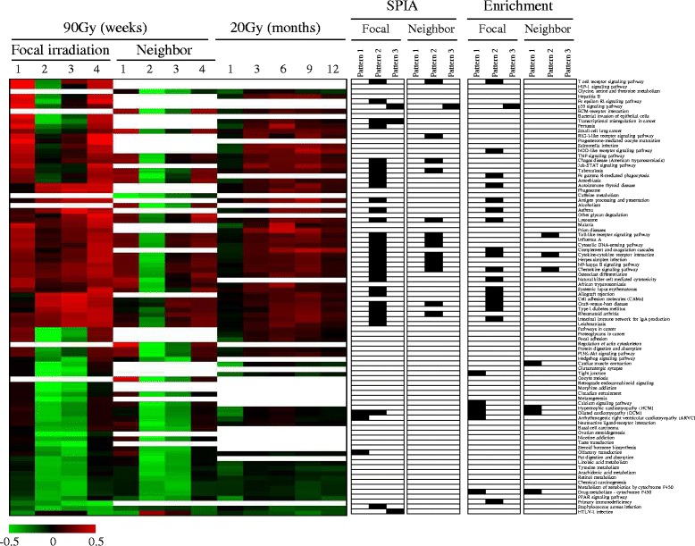
Results: Central areas of the mouse left lung were focally-irradiated (3 mm in diameter) with a single high-dose of radiation (90 Gy). Temporal changes in gene expression in the irradiated and non-irradiated neighboring lung regions were analyzed by microarray. For comparison, the long-term effect (12 months) of 20 Gy radiation on a diffuse region of lung was also measured. The majority of genes were down-regulated in the focally-irradiated lung areas at 2 to 3 weeks after irradiation. This pattern of gene expression was clearly different than gene expression in the diffuse region of lungs exposed to low-dose radiation. Ontological and pathway analyses indicated these down-regulated genes were mainly associated with organ development. Although the number was small, genes that were up-regulated after focal irradiation were associated with immune-related functions. The temporal patterns of gene expression and the associated biological functions were also similar in non-irradiated neighboring lung regions, although statistical significance was greatly reduced when compared with those from focally-irradiated areas of the lung. From network analysis of temporally regulated genes, we identified inter-related modules associated with diverse functions, including organ development and the immune response, in both the focally-irradiated regions and non-irradiated neighboring lung regions.

Conclusions: Focal exposure of lung tissue to high-dose radiation induced expression of genes associated with organ development and the immune response. This pattern of gene expression was also observed in non-irradiated neighboring areas of lung tissue, indicating a global lung response to focal high-dose irradiation.


Figures

Fig. 1
Fig. 1
Fibrotic changes in the control or irradiated lung tissues after focal 90 Gy or 20 Gy irradiation. Mice were sacrificed at the indicated time points after irradiation, and the lungs were immersed in fixation solution. a Representative images of hematoxylin–eosin-stained lung sections from three mice are shown at each time point. The arrows indicate the focally irradiated area. b Lung sections were stained with Masson’s Trichrome stain to visualize blue-colored collagen deposition and quantitative assessments of the degree of collagen deposition were determined using an image J program (*p <0.05 vs non-irradiated control)
Fig. 2
Fig. 2
Temporal gene expression profile induced by radiation in mouse lung. Approximately 6,500 differentially expressed genes with a fold ratio greater than two or less than 0.5 (for up- and down-regulation, respectively) compared to the control in at least one sample were clustered hierarchically. Up-cluster and Down-cluster indicate the two sub-clusters. Columns and rows represent individual samples and genes, respectively. The expression ratio color scale ranges from red (high) to green (low), as indicated by the scale bar with log 2 units
Fig. 3
Fig. 3
Temporal gene expression patterns by focal exposure to high-dose radiation. Temporally altered genes were identified by the Short Time-series Expression Miner (STEM) analysis in (a) focally-irradiated regions and (b) non-irradiated neighboring lung regions. Genes were classified into three patterns (FDR <0.001) in both lung regions. Pattern 1, Pattern 2, and Pattern 3 comprised 4609, 676, and 64 genes, respectively, in (a) focally irradiated regions, and 4893, 218, and 27, respectively, in (b) non-irradiated neighboring lung regions. (c) The number of common genes between the two lung regions was compared according to the three patterns
Fig. 4
Fig. 4
Altered GO terms by focal exposure to high-dose radiation. The network structure among non-redundant GO terms was constructed from all enriched GO terms (FDR <0.01) using the REIVGO program in (a) focally-irradiated regions and (b) non-irradiated neighboring lung regions. The node size and color intensity are proportional to the hierarchical status and statistical significance of each node, respectively. The edge thickness between nodes represents the closeness of the two nodes. c Significantly enriched non-redundant GO terms at each time point were temporally distributed. The columns represent individual samples, while the rows represent statistically significant GO terms (FDR <0.01). The positions of the organ development-related terms and immune-related terms are indicated as bars. A full list of GO terms is depicted in Additional file 6
Fig. 5
Fig. 5
Pathways altered by focal exposure to high-dose radiation. Pathways involved in Pattern 1, Pattern 2, Pattern 3, and all Patterns were analyzed via the Signaling Pathway Impact Analysis (SPIA) program in (a) focally-irradiated regions and (b) non-irradiated neighboring lung regions. The horizontal axis represents pathway over‐representation (PNDE), while the vertical axis indicates pathway perturbation (PPERT). The dotted horizontal and vertical lines represent the corrected thresholds (1 %) of significance (red color for Bonferroni and blue for FDR correction) for each axis value. The red and blue circles at the right of the oblique lines are significant pathways with KEGG ID after the correction (red line for 1 % and blue line for 5 % FDR correction) of the global p-values, PG. PG, representing pathway rank, was calculated from the combined probability of both PNDE and PPERT. The list of pathways for the red circles is shown in Table 3
Fig. 6
Fig. 6
Pathway activities temporally changed by irradiation. Pathway activities were calculated by linearly combining gene expression levels and then were hierarchically clustered. The columns represent individual samples and the rows represent the pathways. The red and green colors reflect high and low activity levels, respectively, as indicated by the scale bar with arbitrary units. Pathways enriched from SPIA and simple enrichment analysis (FDR <0.01) are highlighted in black at the right panel with names
Fig. 7
Fig. 7
Interaction network of genes induced by focal exposure to high-dose radiation. By implementing Reactome FI application, an interaction network was constructed among 6681 and 6799 differentially expressed genes with at least two-fold variation in (a) focally-irradiated regions and in (b) non-irradiated neighboring lung regions, respectively. Both networks from (a) and (b) are composed of a total of 11 modules (as indicated from 0 to 10), each of which is differently colored. The representative GO term associated with each module was obtained from REVIGO program using module genes (FDR <0.01)

References

    1. Timmerman R, Paulus R, Galvin J, Michalski J, Straube W, Bradley J, et al. Stereotactic body radiation therapy for inoperable early stage lung cancer. JAMA. 2010;303:1070–6. doi: 10.1001/jama.2010.261. - DOI - PMC - PubMed
    1. Nagata Y. Stereotactic body radiotherapy for early stage lung cancer. Cancer Res Treat. 2013;45:155–61. doi: 10.4143/crt.2013.45.3.155. - DOI - PMC - PubMed
    1. Mehta V. Radiation pneumonitis and pulmonary fibrosis in non-small-cell lung cancer: pulmonary function, prediction, and prevention. Int J Radiat Oncol Biol Phys. 2005;63:5–24. doi: 10.1016/j.ijrobp.2005.03.047. - DOI - PubMed
    1. Ding NH, Li JJ, Sun LQ. Molecular mechanisms and treatment of radiation-induced lung fibrosis. Curr Drug Targets. 2013;14:1347–56. doi: 10.2174/13894501113149990198. - DOI - PMC - PubMed
    1. Baker R, Han G, Sarangkasiri S, DeMarco M, Turke C, Stevens CW, et al. Clinical and dosimetric predictors of radiation pneumonitis in a large series of patients treated with stereotactic body radiation therapy to the lung. Int J Radiat Oncol Biol Phys. 2013;85:190–5. doi: 10.1016/j.ijrobp.2012.03.041. - DOI - PubMed

Publication types