

Case Reports
doi: 10.1136/bcr-2015-213719.
Necrotic lipoma at the posterior thigh mimicking liposarcoma
Affiliations
- PMID: 26818690
- PMCID: PMC4735368
- DOI: 10.1136/bcr-2015-213719
Item in Clipboard
Case Reports
Necrotic lipoma at the posterior thigh mimicking liposarcoma
BMJ Case Rep.
.
Abstract
A lipoma is one of the most common benign tumours and can develop at any location in the body. Lipomas present characteristic imaging features; hence, they are easy to identify on CT and MRI. However, cases of necrotic lipoma are rarely encountered; therefore, information on the imaging findings of necrotic lipomas is scarce. In the present report, we describe the case of a 63-year-old man with necrotic lipoma in the deep layer of the posterior thigh, which resembled a liposarcoma on imaging. To the best of our knowledge, only a few reports on necrotic lipoma on the extremities have been published.
2016 BMJ Publishing Group Ltd.
Figures
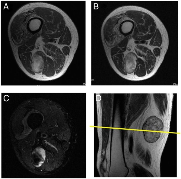
(A) T1-weighted, (B) T2-weighted fat suppression, (C) contrast-enhanced T1-weighted fat suppression preoperative axial view, (D) T1-weighted sagittal view of MRI shows a well-demarcated round lipomatous lesion in the posterior thigh with a non-lipomatous component at the periphery of the tumour. The non-lipomatous component shows good enhancement on gadolinium administration.

(A) Macroscopic picture demonstrating a well-defined yellow mass, which is consistent with necrotic lipoma. (B) On histological examination, the tumour was composed of necrotic adipose tissues. Note the highly degenerative area with focal fibrosis on the left side, which is enhanced on MRI. (C) On higher magnification (square area in B), the adipose tissue in the total necrosis area shows focal cystic degeneration and fibrosis. (D) CD34 immunostaining indicates the vascular channels in the fibrous area.
Similar articles
-
Lipoma or liposarcoma? A cautionary case report.J Plast Reconstr Aesthet Surg. 2012 Jan;65(1):e11-4. doi: 10.1016/j.bjps.2011.08.004. Epub 2011 Aug 23. J Plast Reconstr Aesthet Surg. 2012. PMID: 21865105
-
Imaging of atypical lipomas of the extremities: report of three cases.Skeletal Radiol. 1988;17(7):472-5. doi: 10.1007/BF00364039. Skeletal Radiol. 1988. PMID: 3201273
-
[A clinical study of primary lipoma and liposarcoma of the orbit].Zhonghua Yan Ke Za Zhi. 1992 Nov;28(6):350-1. Zhonghua Yan Ke Za Zhi. 1992. PMID: 1306470 Chinese.
-
MR and CT findings in a case of hibernoma of the thigh extending into the pelvis.Eur Radiol. 1998;8(3):476-8. doi: 10.1007/s003300050419. Eur Radiol. 1998. PMID: 9510590 Review.
-
Atypical lipomatous tumor/well-differentiated liposarcoma of the parotid gland: case report and literature review.Ear Nose Throat J. 2009 Oct;88(10):E10-6. Ear Nose Throat J. 2009. PMID: 19826985 Review.
Cited by
-
Surgical Approach to a Post-traumatic Fat Fracture.Cureus. 2018 Sep 27;10(9):e3378. doi: 10.7759/cureus.3378. Cureus. 2018. PMID: 30510886 Free PMC article.
References
-
- Fletcher CDM, Unnni KK, Mertens F. World Health Organization classification of tumours. Pathology and genetics. Tumours of soft tissue and bone. Lyon: IARC Press, 2014:20–2.
Publication types
MeSH terms
LinkOut - more resources
Full Text Sources
Other Literature Sources