Review
. 2016;14(6):593-609.
doi: 10.2174/1570159x14666151126215358.

Potential Therapeutic Benefits of Maintaining Mitochondrial Health in Peripheral Neuropathies

Affiliations
Review

Potential Therapeutic Benefits of Maintaining Mitochondrial Health in Peripheral Neuropathies

Aparna Areti et al. Curr Neuropharmacol. 2016.

Abstract

Background: Peripheral neuropathies are a group of diseases characterized by malfunctioning of peripheral nervous system. Neuropathic pain, one of the core manifestations of peripheral neuropathy remains as the most severe disabling condition affecting the social and daily routine life of patients suffering from peripheral neuropathy.

Method: The current review is aimed at unfolding the possible role of mitochondrial dysfunction in peripheral nerve damage and to discuss on the probable therapeutic strategies against neuronal mitotoxicity. The article also highlights the therapeutic significance of maintaining a healthy mitochondrial environment in neuronal cells via pharmacological management in context of peripheral neuropathies.

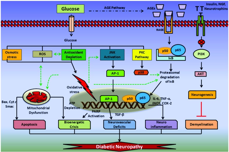
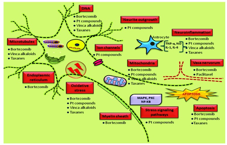
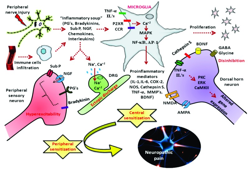
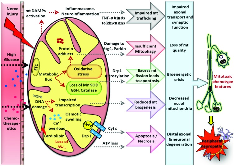
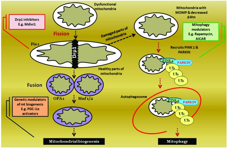
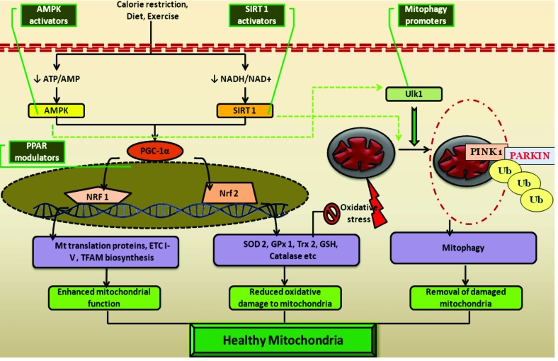
Results: Aberrant cellular signaling coupled with changes in neurotransmission, peripheral and central sensitization are found to be responsible for the pathogenesis of variant toxic neuropathies. Current research reports have indicated the possible involvement of mitochondria mediated redox imbalance as one of the principal causes of neuropathy aetiologies. In addition to imbalance in redox homeostasis, mitochondrial dysfunction is also responsible for alterations in physiological bioenergetic metabolism, apoptosis and autophagy pathways.

Conclusions: In spite of various etiological factors, mitochondrial dysfunction has been found to be a major pathomechanism underlying the neuronal dysfunction associated with peripheral neuropathies. Pharmacological modulation of mitochondria either directly or indirectly is expected to yield therapeutic relief from various primary and secondary mitochondrial diseases.


Figures

Fig. (1)
Fig. (1)
Pathophysiology of Diabetic neuropathy: Glucose induced osmotic stress, reactive oxygen species (ROS) and antioxidant depletion results in the formation of intense oxidative stress inside neuronal cells. Glucose is also found to activate JNK and protein kinase pathway (PKC). JNK activation leads to the facilitation of activator protein-1 (AP-1) directed expression of tumor growth factor-β (TGF-β) and other cytokines, which reduces vascular supply to the nervous tissue. Oxidative damage to mitochondria results in release of pro-apoptotic mediators from the mitochondria and also causes damage to electron transport chain components (ETC). This eventually results in apoptosis and bioenergetic dysfunction associated with DN. Glucose induced advanced glycation end products (AGE) and its further association with receptors for AGE (RAGE) also involved in activation of NF-κB heterodimer and thus releases proinflammatory mediators such as interleukin-6 (IL-6), tumor necrosis factor-α (TNF-α), cyclooxygenase-2 (COX-2) release and thus produces neuroinflammation. Further, lack of insulin, neurotrophic signaling leads to reduced PI3K/AKT mediated neurogeneis and thus may promote demyelination observed in chronic neuropathies.
Fig. (2)
Fig. (2)
Pathogenesis of chemotherapy induced peripheral neuropathy: Multiple sites of peripheral neurons get attacked by various chemotherapeutic agents such as taxane derivatives, vinca alkaloids, Platinum compounds and bortezomib. They adversely affect diverse components at cellular and sub cellular level like ion channels, myelin sheath, DNA, microtubules, mitochondria, endoplasmic reticulum etc. Though these agents cause neurotoxicity by different mechanisms, several mechanisms shared by them in common like oxidative stress, neuroinflammation and mitochondrial dysfunction.
Fig. (3)
Fig. (3)
Pathogenesis of nerve injury induced neuropathic pain: Nerve injury can cause persistent neuropathic pain through pathological cascade of events like release of pain mediators, hyperexcitability in sensory neurons, ectopic discharges, central sensitization and disinhibition. Activation of glial cells in the peripheral nervous system through purinergic and chemokine receptors release inflammatory mediators like (IL-1, IL-6, COX-2, NOS, Cathepsin S, TNF-α, MMP’s, BDNF) through activation of transcription factors like NF-κB, AP-1. These act on central afferent fibers causing long term central sensitization and disinhibition phenomenon. This peripheral and central sensitization occurs as a result of oxidative stress and neuroinflammation generated as a consequence of nerve injury induced glial cell activation.
Fig. (4)
Fig. (4)
Mitotoxicity in peripheral neuropathies: Various pathophysiological insults like hyperglycemic, chemotherapeutic and traumatic injury to the peripheral nerves results in mitochondrial dysfunction through enhanced generation of ROS induced biomolecular damage and bioenergetic crisis. Following the nerve injury accumulation of mitochondria occurs resulting in the release of mtDNA & formyl peptides into circulation which acts as Death associated molecular patterns (DAMP’s). These are recognized by immune cells as foreign bodies and can elicit a local immune/inflammatory response. Interaction between inflammatory mediators and structural proteins involved in mitochondrial trafficking will cause impairment in mitochondrial motility. Oxidative stress induced damage to the mt proteins like Atg4, Parkin etc cause insufficient mitophagy. Excess nitrosative stress also results in excessive mt fission associated with apoptosis. In addition, mtDNA damage impairs its transcription and reduces mitochondrial biogenesis. Ca2+ dyshomeostasis, loss in mitochondrial potential and bioenergetic crisis cause neuronal death via apoptosis/necrosis. All these modifications cause defects in ultra structure, physiology and trafficking of mitochondria resulting in loss of neuronal function producing peripheral neuropathy.
Fig. (5)
Fig. (5)
Regulation of mitochondrial quantity & quality: Mitochondria are dynamic cellular organelles, the quantity and quality of which are maintained through regulated processes of fission, fusion and mitophagy. Fission generally precedes mitophagy. Dysfunctional mitochondria undergoes fission using the help of GTPase proteins such as Fis1, dynamin related protein (Drp1). Damaged parts of mitochondria will further undergo recycling using a catabolic mitophagy process. Such damaged mitochondria undergoes PINK1, PARKIN recruitment mediated ubiquitination and then accumulation in autophagosomes. Healthy parts of mitochondria in turn undergo fusion process using the help of proteins such as OPA1, Mnf 1/2. All of these stages of mitochondrial regulation can be pharmacologically modulated pertaining to the catering needs of cells. Excessive mitochondrial fission can be inhibited by using Drp1 inhibitor (e.g. Mdivi1). Mitophagy can be enhanced by various cellular transcription modulators (e.g. mTOR inhibitors).
Fig. (6)
Fig. (6)
Cellular manipulators of mitochondria and their pharmacological modulation: Cellular energetic sensors such as adenosine monophosphate kinase (AMPK), silent information regulator of transcription homologue type 1 (SIRT1) activated in response to nutrient starvation and at the periods of high metabolic demand. These energetic sensors activate PPAR-γ coactivator-1α (PGC-1α) by direct phosphorylation. The activated PGC-1α in turn regulates mitochondrial biosynthesis through transcriptional facilitation of nuclear respiratory factor-1 (NRF-1) and reduces cellular oxidative damage by enhancing the transcription of antioxidant response element (ARE) of genome through activation of nuclear erythroid factor-1(NEF-1) related facor-2 (Nrf-2). Further AMPK and SIRT1 are also found to promote mitophagy either directly activating Unc-51 like kinase 1 (Ulk1) or indirectly through mTOR inhibition. All these resulting events facilitate the mitochondrial function, reduce damage to it and recycle the constituents if damage occurs, the characteristic features of a healthy mitochondrial phenotype. The mitochondrial transcription facilitator PGC-1α can be promoted by using peroxisome proliferator activated receptor modulators (e.g. Glitazones). Similarly, PGC-1α mediated mitochondrial functions can also be indirectly activated through specific AMPK and SIRT1 activators. Direct pharmacological promoters of mitophagy can also be useful in maintaining a healthy population of mitochondria (e.g. mTOR inhibitors).

References

    1. Ganesh Yerra V., Negi G., Sharma S.S., Kumar A. Potential therapeutic effects of the simultaneous targeting of the Nrf2 and NF-κB pathways in diabetic neuropathy. Redox Biol. 2013;1:394–397. [http://dx.doi.org/10.1016/j.redox.2013.07.005]. [PMID: 24024177]. - PMC - PubMed
    1. Baron R., Binder A., Wasner G. Neuropathic pain: diagnosis, pathophysiological mechanisms, and treatment. Lancet Neurol. 2010;9(8):807–819. [http://dx.doi.org/10.1016/S1474-4422(10) 70143-5]. [PMID: 20650402]. - PubMed
    1. Weisberg L.A., Garcia C., Strub R.L. Essentials of clinical neurology. St Louis: Mosby; 1996. Diseases of the peripheral nerves and motor neurons. pp. 458–494.
    1. Saporta M.A., Shy M.E. Inherited peripheral neuropathies. Neurol. Clin. 2013;31(2):597–619. [http://dx.doi.org/10.1016/ j.ncl.2013.01.009]. [PMID: 23642725]. - PMC - PubMed
    1. Carelli V., Ross-Cisneros F.N., Sadun A.A. Mitochondrial dysfunction as a cause of optic neuropathies. Prog. Retin. Eye Res. 2004;23(1):53–89. [http://dx.doi.org/10.1016/j.preteyeres.2003.10. 003]. [PMID: 14766317]. - PubMed

MeSH terms