

Molecular evolution of gas cavity in [NiFeSe] hydrogenases resurrected in silico
- PMID: 26818780
- PMCID: PMC4730141
- DOI: 10.1038/srep19742
Molecular evolution of gas cavity in [NiFeSe] hydrogenases resurrected in silico
Abstract
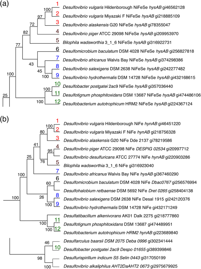
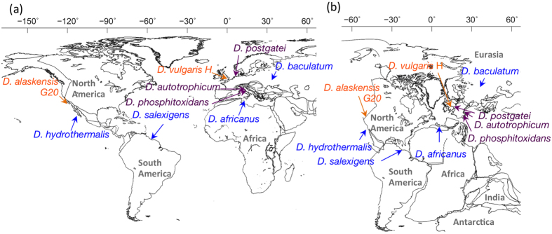
Oxygen tolerance of selenium-containing [NiFeSe] hydrogenases (Hases) is attributable to the high reducing power of the selenocysteine residue, which sustains the bimetallic Ni-Fe catalytic center in the large subunit. Genes encoding [NiFeSe] Hases are inherited by few sulphate-reducing δ-proteobacteria globally distributed under various anoxic conditions. Ancestral sequences of [NiFeSe] Hases were elucidated and their three-dimensional structures were recreated in silico using homology modelling and molecular dynamic simulation, which suggested that deep gas channels gradually developed in [NiFeSe] Hases under absolute anaerobic conditions, whereas the enzyme remained as a sealed edifice under environmental conditions of a higher oxygen exposure risk. The development of a gas cavity appears to be driven by non-synonymous mutations, which cause subtle conformational changes locally and distantly, even including highly conserved sequence regions.
Figures


Similar articles
-
Hydrogenases in Desulfovibrio vulgaris Hildenborough: structural and physiologic characterisation of the membrane-bound [NiFeSe] hydrogenase.J Biol Inorg Chem. 2005 Oct;10(6):667-82. doi: 10.1007/s00775-005-0022-4. Epub 2005 Nov 2. J Biol Inorg Chem. 2005. PMID: 16187073
-
[NiFeSe]-hydrogenase chemistry.Acc Chem Res. 2015 Nov 17;48(11):2858-65. doi: 10.1021/acs.accounts.5b00326. Epub 2015 Oct 21. Acc Chem Res. 2015. PMID: 26488197
-
The three-dimensional structure of [NiFeSe] hydrogenase from Desulfovibrio vulgaris Hildenborough: a hydrogenase without a bridging ligand in the active site in its oxidised, "as-isolated" state.J Mol Biol. 2010 Mar 5;396(4):893-907. doi: 10.1016/j.jmb.2009.12.013. Epub 2009 Dec 21. J Mol Biol. 2010. PMID: 20026074
-
Structure and function of [NiFe] hydrogenases.J Biochem. 2016 Nov;160(5):251-258. doi: 10.1093/jb/mvw048. Epub 2016 Aug 4. J Biochem. 2016. PMID: 27493211 Review.
-
Hydrogenases and H(+)-reduction in primary energy conservation.Results Probl Cell Differ. 2008;45:223-52. doi: 10.1007/400_2006_027. Results Probl Cell Differ. 2008. PMID: 18500479 Review.
References
-
- Vignais P. M. & Billoud B. Occurrence, classification, and biological function of hydrogenases: An overview. Chem. Rev. 107, 4206–4272 (2007). - PubMed
-
- Böck A. et al. Selenocysteine: the 21st amino acid. Mol. Microbiol. 5, 515–520 (1991). - PubMed
-
- Teixeira M. et al. Redox properties and activity studies on a nickel-containing hydrogenase isolated from a halophilic sulfate reducer Desulfovibrio salexigens. Biochimie. 68, 75–84 (1986). - PubMed
-
- Valente F. M. et al. Hydrogenases in Desulfovibrio vulgaris Hildenborough: structural and physiologic characterization of the membrane-bound [NiFeSe]-hydrogenase. J. Biol. Inorg. Chem. 10, 667–682 (2005). - PubMed
-
- Nonaka K., Nguyen N. T., Yoon K. S. & Ogo S. Novel H2-oxidizing [NiFeSe]-hydrogenase from Desulfovibrio vulgaris Miyazaki F. J. Biosci. Bioeng. 115, 366–371 (2013). - PubMed
Publication types
MeSH terms
Substances
LinkOut - more resources
Full Text Sources
Other Literature Sources
Molecular Biology Databases
