

Microarchitecture and Peripheral BMD are Impaired in Postmenopausal White Women With Fracture Independently of Total Hip T-Score: An International Multicenter Study
- PMID: 26818785
- PMCID: PMC4891284
- DOI: 10.1002/jbmr.2796
Microarchitecture and Peripheral BMD are Impaired in Postmenopausal White Women With Fracture Independently of Total Hip T-Score: An International Multicenter Study
Abstract
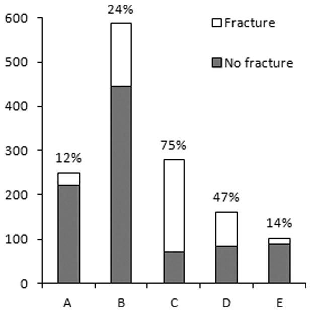
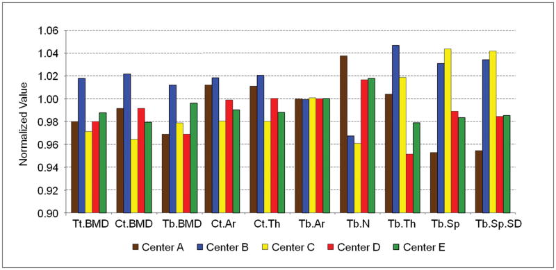
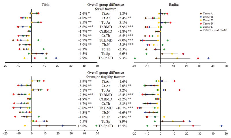
Because single-center studies have reported conflicting associations between microarchitecture and fracture prevalence, we included high-resolution peripheral quantitative computed tomography (HR-pQCT) data from five centers worldwide into a large multicenter analysis of postmenopausal women with and without fracture. Volumetric BMD (vBMD) and microarchitecture were assessed at the distal radius and tibia in 1379 white postmenopausal women (age 67 ± 8 years); 470 (34%) had at least one fracture including 349 with a major fragility fracture. Age, height, weight, and total hip T-score differed across centers and were employed as covariates in analyses. Women with fracture had higher BMI, were older, and had lower total hip T-score, but lumbar spine T-score was similar between groups. At the radius, total and trabecular vBMD and cortical thickness were significantly lower in fractured women in three out of five centers, and trabecular number in two centers. Similar results were found at the tibia. When data from five centers were combined, however, women with fracture had significantly lower total, trabecular, and cortical vBMD (2% to 7%), lower trabecular number (4% to 5%), and thinner cortices (5% to 6%) than women without fracture after adjustment for covariates. Results were similar at the radius and tibia. Similar results were observed with analysis restricted to major fragility fracture, vertebral and hip fractures, and peripheral fracture (at the radius). When focusing on osteopenic women, each SD decrease of total and trabecular vBMD was associated with a significantly increased risk of major fragility fracture (OR = 1.55 to 1.88, p < 0.01) after adjustment for covariates. Moreover, trabecular architecture modestly improved fracture discrimination beyond peripheral total vBMD. In conclusion, we observed differences by center in the magnitude of fracture/nonfracture differences at both the distal radius and tibia. However, when data were pooled across centers and the sample size increased, we observed significant and consistent deficits in vBMD and microarchitecture independent of total hip T-score in all postmenopausal white women with fracture and in the subgroup of osteopenic women, compared to women who never had a fracture. © 2016 American Society for Bone and Mineral Research.
Keywords: BONE MICROSTRUCTURE; FRACTURE RISK; HR-PQCT; MULTICENTER STUDIES; OSTEOPOROSIS.
© 2016 American Society for Bone and Mineral Research.
Conflict of interest statement
All authors state that they have no conflicts of interest.
Figures
Similar articles
-
In vivo assessment of trabecular bone microarchitecture by high-resolution peripheral quantitative computed tomography.J Clin Endocrinol Metab. 2005 Dec;90(12):6508-15. doi: 10.1210/jc.2005-1258. Epub 2005 Sep 27. J Clin Endocrinol Metab. 2005. PMID: 16189253
-
Deterioration of trabecular plate-rod and cortical microarchitecture and reduced bone stiffness at distal radius and tibia in postmenopausal women with vertebral fractures.Bone. 2016 Jul;88:39-46. doi: 10.1016/j.bone.2016.04.003. Epub 2016 Apr 12. Bone. 2016. PMID: 27083398 Free PMC article.
-
Impaired trabecular and cortical microarchitecture in daughters of women with osteoporotic fracture: the MODAM study.Osteoporos Int. 2013 Jun;24(6):1881-9. doi: 10.1007/s00198-012-2223-3. Epub 2012 Nov 23. Osteoporos Int. 2013. PMID: 23179577
-
Mortality and morbidity in patients with osteogenesis imperfecta in Denmark.Dan Med J. 2018 Apr;65(4):B5454. Dan Med J. 2018. PMID: 29619932 Review.
-
Best Performance Parameters of HR-pQCT to Predict Fragility Fracture: Systematic Review and Meta-Analysis.J Bone Miner Res. 2021 Dec;36(12):2381-2398. doi: 10.1002/jbmr.4449. Epub 2021 Oct 18. J Bone Miner Res. 2021. PMID: 34585784 Free PMC article.
Cited by
-
Differences between bone health parameters in adults with acromegaly and growth hormone deficiency: A systematic review.Best Pract Res Clin Endocrinol Metab. 2023 Dec;37(6):101824. doi: 10.1016/j.beem.2023.101824. Epub 2023 Sep 28. Best Pract Res Clin Endocrinol Metab. 2023. PMID: 37798201 Free PMC article.
-
The reproducibility of measuring trabecular bone parameters using a commercially available high-resolution magnetic resonance imaging approach: A pilot study.Bone Rep. 2018 Apr 26;8:180-186. doi: 10.1016/j.bonr.2018.04.006. eCollection 2018 Jun. Bone Rep. 2018. PMID: 29955637 Free PMC article.
-
Gene Variants and Bisphosphonates Treatment in Pregnancy and Lactation-Associated Osteoporosis (PLO): A Retrospective Study of 22 Chinese Patients.Calcif Tissue Int. 2025 May 6;116(1):70. doi: 10.1007/s00223-025-01381-x. Calcif Tissue Int. 2025. PMID: 40327138
-
Hip Fractures in Older Adults Are Associated With the Low Density Bone Phenotype and Heterogeneous Deterioration of Bone Microarchitecture.J Bone Miner Res. 2022 Oct;37(10):1963-1972. doi: 10.1002/jbmr.4663. Epub 2022 Aug 22. J Bone Miner Res. 2022. PMID: 35895080 Free PMC article.
-
Persistent changes in calcium-regulating hormones and bone turnover markers in living kidney donors more than 20 years after donation.JBMR Plus. 2024 May 13;8(7):ziae067. doi: 10.1093/jbmrpl/ziae067. eCollection 2024 Jul. JBMR Plus. 2024. PMID: 38868597 Free PMC article.
References
-
- Boutroy S, Bouxsein ML, Munoz F, Delmas PD. In vivo assessment of trabecular bone microarchitecture by high-resolution peripheral quantitative computed tomography. J Clin Endocrinol Metab. 2005;90(12):6508–15. - PubMed
-
- Boutroy S, Van Rietbergen B, Sornay-Rendu E, Munoz F, Bouxsein ML, Delmas PD. Finite element analysis based on in vivo HR-pQCT images of the distal radius is associated with wrist fracture in postmenopausal women. J Bone Miner Res. 2008;23(3):392–9. - PubMed
-
- Sornay-Rendu E, Boutroy S, Munoz F, Delmas PD. Alterations of cortical and trabecular architecture are associated with fractures in postmenopausal women, partially independent of decreased BMD measured by DXA: the OFELY study. J Bone Miner Res. 2007;22(3):425–33. - PubMed
-
- Sornay-Rendu E, Cabrera-Bravo JL, Boutroy S, Munoz F, Delmas PD. Severity of vertebral fractures is associated with alterations of cortical architecture in postmenopausal women. J Bone Miner Res. 2009;24(4):737–43. - PubMed
-
- Vico L, Zouch M, Amirouche A, Frere D, Laroche N, Koller B, et al. High-resolution pQCT analysis at the distal radius and tibia discriminates patients with recent wrist and femoral neck fractures. J Bone Miner Res. 2008;23(11):1741–50. - PubMed
Publication types
MeSH terms
Grants and funding
LinkOut - more resources
Full Text Sources
Other Literature Sources
Medical
