

Overexpression of HMGA2 promotes tongue cancer metastasis through EMT pathway
- PMID: 26818837
- PMCID: PMC4730598
- DOI: 10.1186/s12967-016-0777-0
Overexpression of HMGA2 promotes tongue cancer metastasis through EMT pathway
Abstract
Background: Metastasis to long distance organs is the main reason leading to morality of tongue squamous cell carcinoma (TSCC); however, the molecular mechanisms are still unknown. High mobility group AT-hook 2 (HMGA2) is highly expressed in multiple metastatic carcinomas, in which it contributes to cancer progression, metastasis and poor prognosis by upregulating Snail expression and inducing epithelial mesenchymal transition (EMT). This study focuses on investigating the role and mechanism of regulation of HMGA2 in the metastasis of TSCC.
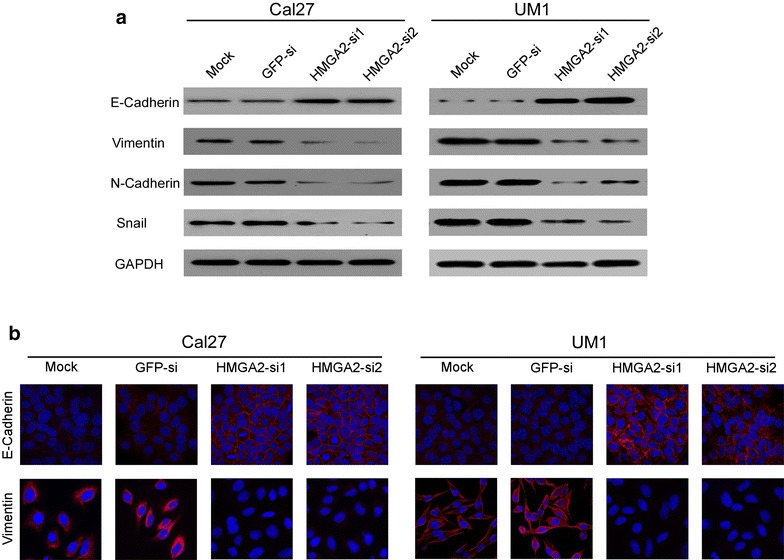
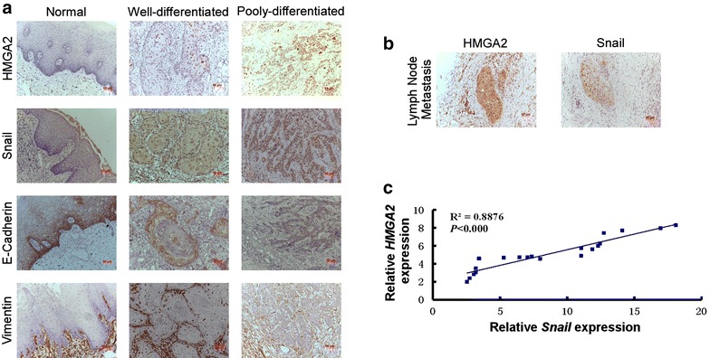
Methods: HMGA2 mRNA and protein expression were examined in TSCC specimens by quantitative real-time polymerase chain reaction, western blotting and immunohistochemistry (IHC). Western blotting, IHC and immunofluorescence were also used to measure the expression and localization of EMT marker E-Cadherin and Vimentin both in TSCC cells and tissues. Knockdown assay was performed in vitro in TSCC cell lines using small interfering RNAs and the functional assay was carried out to determine the role of HMGA2 in TSCC cell migration and invasion.
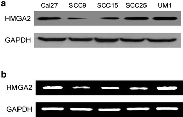
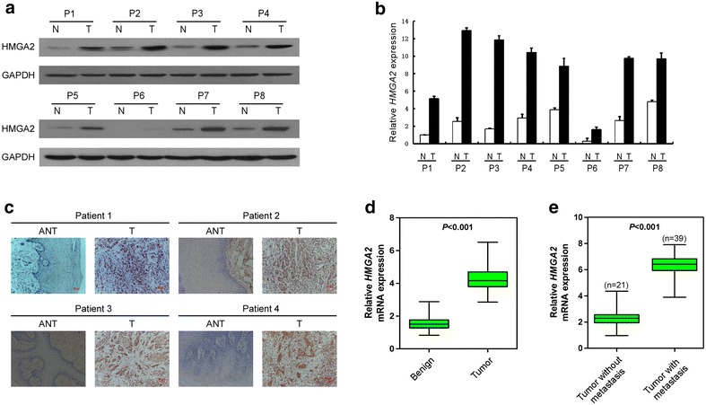
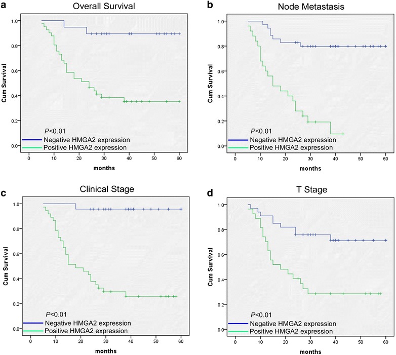
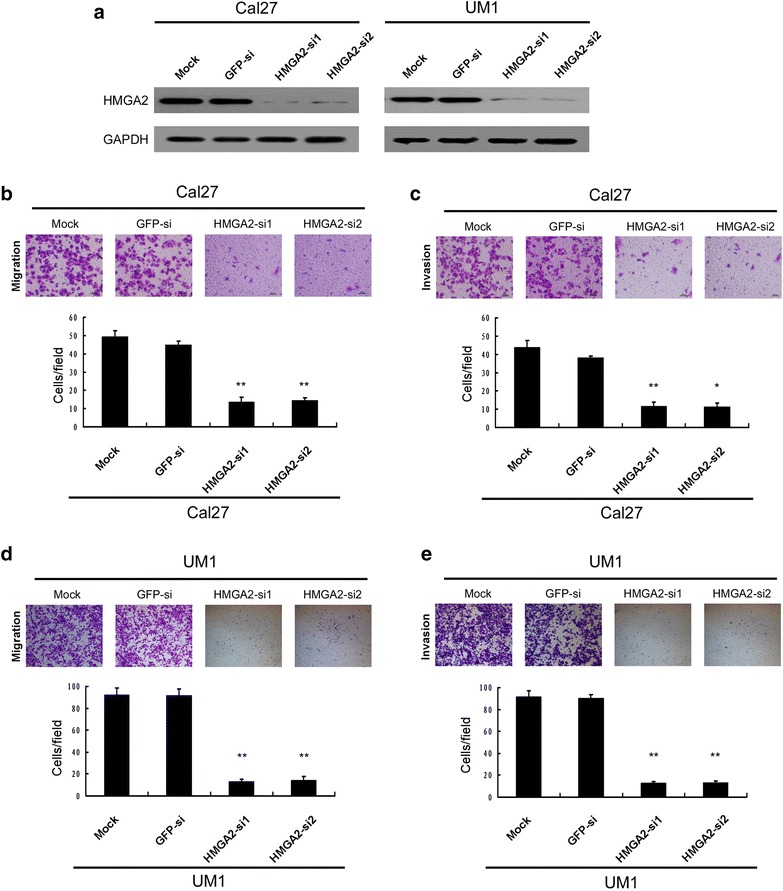
Results: TSCC mRNA and protein expression were significantly up-regulated in tumor tissues when compared to adjacent non-tumor tissues, and the overexpression of HMGA2 was closely correlated with lymph nodes metastasis. Clinicopathological analysis indicated that HMGA2 expression was associated with clinical stage (P = 0.001), lymph node metastasis (P = 0.000), histological differentiation (P = 0.002) and survival (P = 0.000). Silencing the HMGA2 expression in Cal27 and UM1 resulted in the inhibition of cell migration and invasion, meanwhile down-regulation of HMGA2 impaired the phenotype of EMT in TSCC cell lines and tissues. The Multivariate survival analysis indicates that HMGA2 can be an independent prognosis biomarker in TSCC.
Conclusion: Our findings demonstrate that HMGA2 promotes TSCC invasion and metastasis; additionally, HMGA2 is an independent prognostic factor which implied that HMGA2 can be a biomarker both for prognosis and therapeutic target of TSCC.
Figures
Similar articles
-
HMGA2 is associated with the aggressiveness of tongue squamous cell carcinoma.Oral Dis. 2017 Mar;23(2):255-264. doi: 10.1111/odi.12608. Epub 2016 Dec 23. Oral Dis. 2017. PMID: 27809392
-
Wnt7a predicts poor prognosis, and contributes to growth and metastasis in tongue squamous cell carcinoma.Oncol Rep. 2019 Mar;41(3):1749-1758. doi: 10.3892/or.2019.6974. Epub 2019 Jan 22. Oncol Rep. 2019. PMID: 30747225
-
PDGF-D/PDGFRβ promotes tongue squamous carcinoma cell (TSCC) progression via activating p38/AKT/ERK/EMT signal pathway.Biochem Biophys Res Commun. 2016 Sep 16;478(2):845-51. doi: 10.1016/j.bbrc.2016.08.035. Epub 2016 Aug 6. Biochem Biophys Res Commun. 2016. PMID: 27507215
-
MicroRNA expression and its implications for diagnosis and therapy of tongue squamous cell carcinoma.J Cell Mol Med. 2016 Jan;20(1):10-6. doi: 10.1111/jcmm.12650. Epub 2015 Oct 24. J Cell Mol Med. 2016. PMID: 26498914 Free PMC article. Review.
-
MicroRNAs in human tongue squamous cell carcinoma: From pathogenesis to therapeutic implications.Oral Oncol. 2017 Apr;67:124-130. doi: 10.1016/j.oraloncology.2017.02.015. Epub 2017 Feb 27. Oral Oncol. 2017. PMID: 28351566 Review.
Cited by
-
Potential Utility of Cell Free High Mobility Group AT-hook 2 (HMGA2) as a Prognostic Biomarker in Liquid Biopsies of Oral Squamous Cell Carcinoma.Asian Pac J Cancer Prev. 2021 Feb 1;22(2):407-412. doi: 10.31557/APJCP.2021.22.2.407. Asian Pac J Cancer Prev. 2021. PMID: 33639654 Free PMC article.
-
Expression and significant roles of the long non-coding RNA CASC19/miR-491-5p/HMGA2 axis in the development of gastric cancer.World J Gastrointest Oncol. 2024 Aug 15;16(8):3559-3584. doi: 10.4251/wjgo.v16.i8.3559. World J Gastrointest Oncol. 2024. PMID: 39171190 Free PMC article.
-
Long non-coding RNA HIT000218960 is associated with poor prognosis in patients with gastric cancer.Exp Ther Med. 2021 Jul;22(1):694. doi: 10.3892/etm.2021.10126. Epub 2021 May 2. Exp Ther Med. 2021. PMID: 34055048 Free PMC article.
-
Peptide Sequence Influence on the Conformational Dynamics and DNA binding of the Intrinsically Disordered AT-Hook 3 Peptide.Sci Rep. 2018 Jul 17;8(1):10783. doi: 10.1038/s41598-018-28956-z. Sci Rep. 2018. PMID: 30018295 Free PMC article.
-
A review of the most promising biomarkers for early diagnosis and prognosis prediction of tongue squamous cell carcinoma.Br J Cancer. 2018 Sep;119(6):724-736. doi: 10.1038/s41416-018-0233-4. Epub 2018 Aug 21. Br J Cancer. 2018. PMID: 30131545 Free PMC article. Review.
References
Publication types
MeSH terms
Substances
LinkOut - more resources
Full Text Sources
Other Literature Sources
Research Materials
