

Gingival fibromatosis: clinical, molecular and therapeutic issues
- PMID: 26818898
- PMCID: PMC4729029
- DOI: 10.1186/s13023-016-0395-1
Gingival fibromatosis: clinical, molecular and therapeutic issues
Abstract
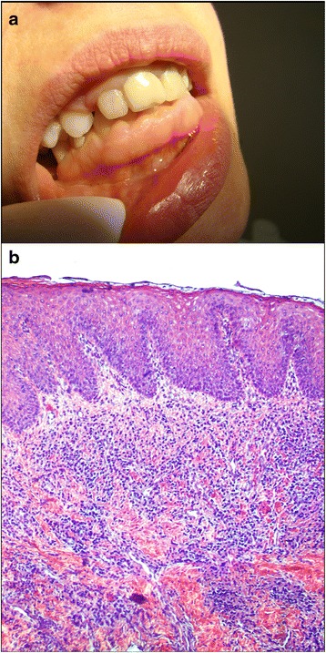
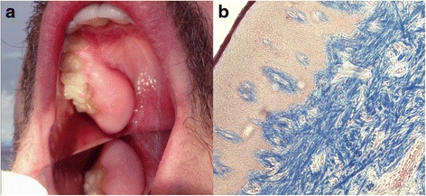
Gingival fibromatosis is a rare and heterogeneous group of disorders that develop as slowly progressive, local or diffuse enlargements within marginal and attached gingiva or interdental papilla. In severe cases, the excess tissue may cover the crowns of the teeth, thus causing functional, esthetic, and periodontal problems, such as bone loss and bleeding, due to the presence of pseudopockets and plaque accumulation. It affects both genders equally. Hereditary, drug-induced, and idiopathic gingival overgrowth have been reported. Hereditary gingival fibromatosis can occur as an isolated condition or as part of a genetic syndrome. The pathologic manifestation of gingival fibromatosis comprises excessive accumulation of extracellular matrix proteins, of which collagen type I is the most prominent example. Mutation in the Son-of-Sevenless-1 gene has been suggested as one possible etiological cause of isolated (non-syndromic) hereditary gingival fibromatosis, but mutations in other genes are also likely to be involved, given the heterogeneity of this condition. The most attractive concept of mechanism for drug-induced gingival overgrowth is epithelial-to-mesenchymal transition, a process in which interactions between gingival cells and the extracellular matrix are weakened as epithelial cells transdifferentiate into fibrogenic fibroblast-like cells. The diagnosis is mainly made on the basis of the patient's history and clinical features, and on histopathological evaluation of affected gingiva. Early diagnosis is important, mostly to exclude oral malignancy. Differential diagnosis comprises all pathologies in the mouth with excessive gingival overgrowth. Hereditary gingival fibromatosis may present as an autosomal-dominant or less commonly autosomal-recessive mode of inheritance. If a systemic disease or syndrome is suspected, the patient is directed to a geneticist for additional clinical examination and specialized diagnostic tests. Treatments vary according to the type of overgrowth and the extent of disease progression, thus, scaling of teeth is sufficient in mild cases, while in severe cases surgical intervention is required. Prognosis is precarious and the risk of recurrence exists.
Figures

References
-
- Pappachan B, Narayan JV, Nayak A. Idiopathic gingival fibromatosis: A neglected case. Indian J Radiol Imaging. 2002;12:335–38.
-
- Hereditary gingival fibromatosis. Orphanet. January 2013. http://www.orpha.net/consor/cgi-bin/Disease_Search.php?lng=EN&data_id=1955. (Accessed 09/09/2015).
-
- Gingival fibromatosis – facial dysmorphism. Orphanet. 2010. http://www.orpha.net/consor/cgi-bin/Disease_Search.php?lng=EN&data_id=1956. (Accessed 15/09/2015).
Publication types
MeSH terms
LinkOut - more resources
Full Text Sources
Other Literature Sources
