. 2016 Jul 5;19(7):pyw007.
doi: 10.1093/ijnp/pyw007. Print 2016 Jul.

Maternal Immune Activation Disrupts Dopamine System in the Offspring

Affiliations

Maternal Immune Activation Disrupts Dopamine System in the Offspring

Antonio Luchicchi et al. Int J Neuropsychopharmacol. .

Abstract

Background: In utero exposure to maternal viral infections is associated with a higher incidence of psychiatric disorders with a supposed neurodevelopmental origin, including schizophrenia. Hence, immune response factors exert a negative impact on brain maturation that predisposes the offspring to the emergence of pathological phenotypes later in life. Although ventral tegmental area dopamine neurons and their target regions play essential roles in the pathophysiology of psychoses, it remains to be fully elucidated how dopamine activity and functionality are disrupted in maternal immune activation models of schizophrenia.

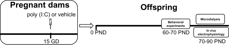
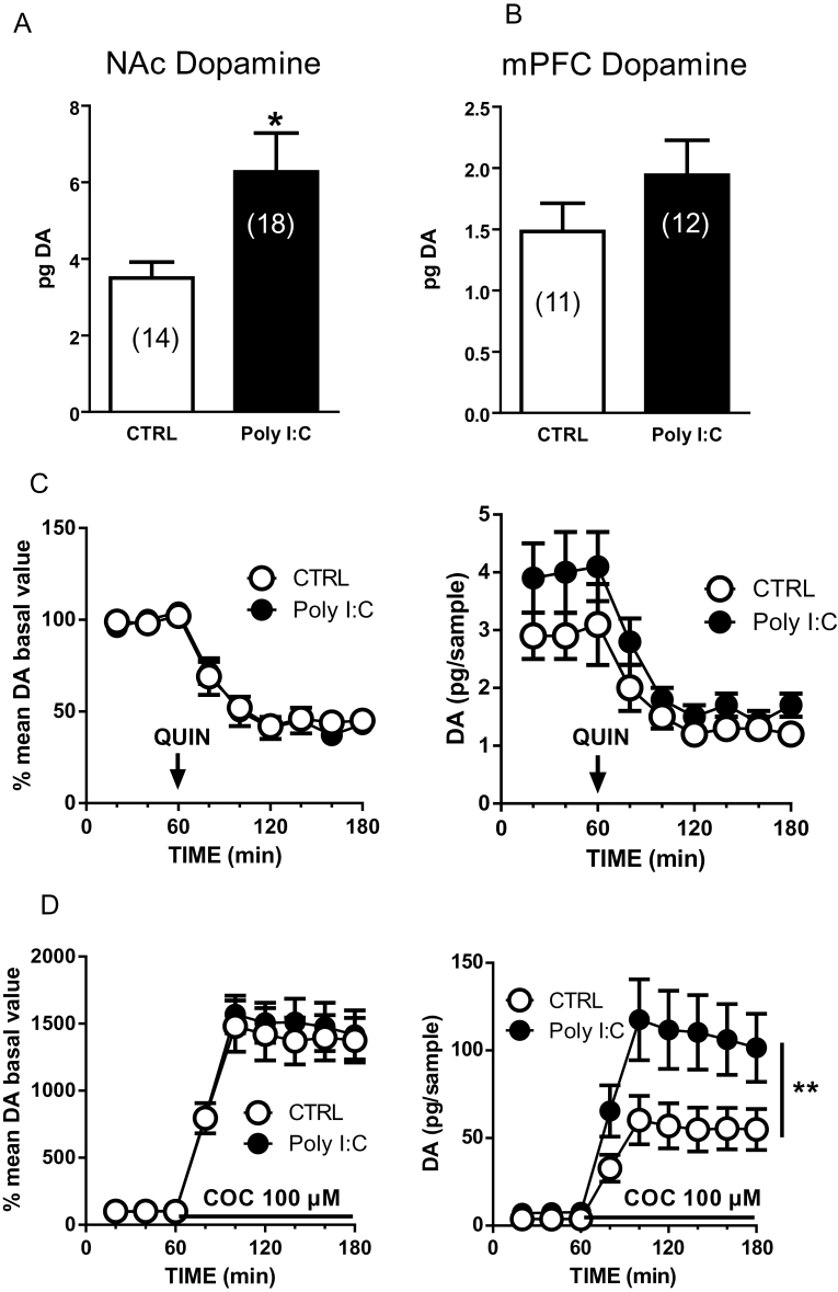
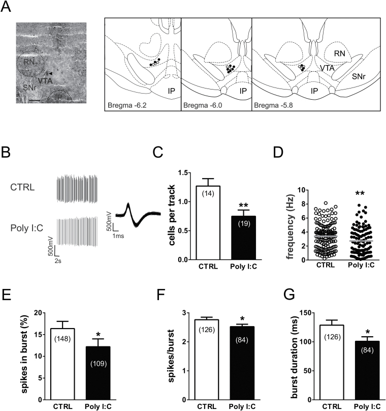
Methods: Here, we used an immune-mediated neurodevelopmental disruption model based on prenatal administration of the polyriboinosinic-polyribocytidilic acid in rats, which mimics a viral infection and recapitulates behavioral abnormalities relevant to psychiatric disorders in the offspring. Extracellular dopamine levels were measured by brain microdialysis in both the nucleus accumbens shell and the medial prefrontal cortex, whereas dopamine neurons in ventral tegmental area were studied by in vivo electrophysiology.

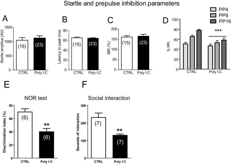
Results: Polyriboinosinic-polyribocytidilic acid-treated animals, at adulthood, displayed deficits in sensorimotor gating, memory, and social interaction and increased baseline extracellular dopamine levels in the nucleus accumbens, but not in the prefrontal cortex. In polyriboinosinic-polyribocytidilic acid rats, dopamine neurons showed reduced spontaneously firing rate and population activity.

Conclusions: These results confirm that maternal immune activation severely impairs dopamine system and that the polyriboinosinic-polyribocytidilic acid model can be considered a proper animal model of a psychiatric condition that fulfills a multidimensional set of validity criteria predictive of a human pathology.

Keywords: Schizophrenia; dopamine; electrophysiology; microdialysis.


Figures

Figure 1.
Figure 1.
Schematic representation of the experimental protocol. Polyriboinosinic-polyribocytidilic acid [poly(I:C)] treatment during pregnancy consisted in a single i.v. injection of poly(I:C) (4mg/kg) or vehicle (sterile pyrogen-free saline) at the 15th gestational day (GD). Behavioral experiments were performed between postnatal day (PND) 60 and 70, whereas in vivo electrophysiology and microdialysis between 70 and 90 PND.
Figure 2.
Figure 2.
Behavioral abnormalities in polyriboinosinic-polyribocytidilic acid [poly(I:C)] offspring. Effects of poly(I:C) on startle reflex (A-C) and prepulse inhibition (PPI) (D) parameters. Graphs show that offspring from poly(I:C)-treated mothers exhibited impairments in PPI but not in startle indices. Values are expressed as mean±SEM. ***P<.001 vs rats treated with vehicle (main effect of treatment). Prepulses are indicated by the intensity corresponding to decibels above background noise. AU, arbitrary units; %IBR, percent inter-block ratio; ms, milliseconds. For further details, see text. (E) Graphs show that offspring of poly(I:C)-treated mothers exhibited reduced discrimination index toward a novel object and (F) reduced social interactions. Data are expressed as means±SEM. *P<.01 vs rats treated with vehicle (CTRL group).
Figure 3.
Figure 3.
Enhanced extracellular dopamine (DA) levels in the nucleus accumbens (NAc) shell of polyriboinosinic-polyribocytidilic acid [poly(I:C)]-treated rats and effects of quinpirole and cocaine. Extracellular dopamine concentrations in the NAc shell (A) and medial prefrontal cortex (mPFC) (B) of control (CTRL) and poly(I:C) rats. (C) These graphs show the effect of systemically administered quinpirole (QUIN, 0.2mg/kg, s.c., n=5–6) on extracellular dopamine levels in the NAc shell expressed as percent of baseline (left) or in absolute values (pg/sample, right). (D) Locally perfused cocaine (COC, 100 µM, n=9–12) enhances extracellular dopamine levels in the NAc shell. Data are expressed as percent of baseline (left) or in absolute values (pg/sample, right). The arrow represents the time of quinpirole administration, whereas the solid line represents the time of cocaine perfusion. Values are the mean±SEM and are expressed as pg/20 µL dialysate. *P<.05, unpaired t test with Welch’s correction.
Figure 4.
Figure 4.
Prenatal polyriboinosinic-polyribocytidilic acid [poly(I:C)] treatment dysregulates dopamine neuron firing activity in adulthood. (A) Example of a recording location for a ventral tegmental area (VTA) dopamine neuron in a poly(I:C)-treated rat (the triangle indicates the pontamine sky blue dye). The diagrams at right show samples of localizations of recording sites in poly(I:C) rats (black dots) and controls (white dots) as verified by histological sections. IP, interpeduncular nucleus; RN, red nucleus; SNr, substantia nigra pars reticulata. Scale bar = 0.5mm. (B) This panel shows traces illustrating representative extracellular recordings of a putative dopamine neuron in the VTA of anesthetized rats belonging to the control group (CTRL, above) and poly(I:C) group (below). Dopamine neurons recorded from poly(I:C) rats typically show slower firing activity and a reduced bursting compared with CTRL. The left trace shows the typical broad spike waveform of a dopamine neuron. (C) The bar graph shows the number of spontaneously active VTA dopamine neurons, which was different between the experimental groups. The scatter plot in (D) displays individual dopamine neuron firing rate in CTRL and poly(I:C) rats. The horizontal lines represent average values that are significantly different between the 2 groups. Graph histograms represent the percentage of spikes in bursts (E), the mean number of spikes per each burst (F), and the mean burst duration (G). Data are expressed as percentage or mean±SEM. *P<.05, **P<.01.

References

    1. Abi-Dargham A, Rodenhiser J, Printz D, Zea-Ponce Y, Gil R, Kegeles LS, Weiss R, Cooper TB, Mann JJ, Van Heertum RL, Gorman JM, Laruelle M. (2000) Increased baseline occupancy of D2 receptors by dopamine in schizophrenia. Proc Natl Acad Sci U S A 97:8104–8109. - PMC - PubMed
    1. Anzalone A, Lizardi-Ortiz JE, Ramos M, De Mei C, Hopf FW, Iaccarino C, Halbout B, Jacobsen J, Kinoshita C, Welter M, Caron MG, Bonci A, Sulzer D, Borrelli E. (2012) Dual control of dopamine synthesis and release by presynaptic and postsynaptic dopamine D2 receptors. J Neurosci 32:9023–9034. - PMC - PubMed
    1. Atladottir HO, Thorsen P, Ostergaard L, Schendel DE, Lemcke S, Abdallah M, Parner ET. (2010) Maternal infection requiring hospitalization during pregnancy and autism spectrum disorders. J Autism Dev Disord 40:1423–1430. - PubMed
    1. Baharnoori M, Bhardwaj SK, Srivastava LK. (2013) Effect of maternal lipopolysaccharide administration on the development of dopaminergic receptors and transporter in the rat offspring. PloS one 8:e54439. - PMC - PubMed
    1. Boksa P. (2010) Effects of prenatal infection on brain development and behavior: a review of findings from animal models. Brain Behav Immun 24:881–897. - PubMed

Publication types

MeSH terms