. 2016 Feb 2;7(5):6294-313.
doi: 10.18632/oncotarget.7034.

PDIP46 (DNA polymerase δ interacting protein 46) is an activating factor for human DNA polymerase δ

Affiliations

PDIP46 (DNA polymerase δ interacting protein 46) is an activating factor for human DNA polymerase δ

Xiaoxiao Wang et al. Oncotarget. .

Abstract

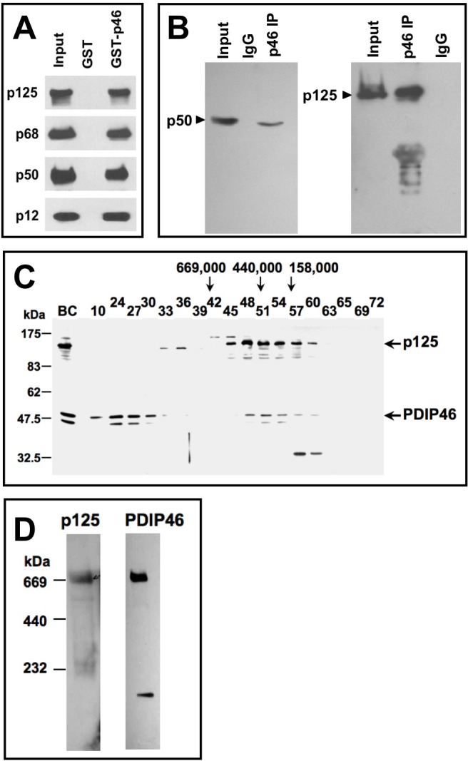
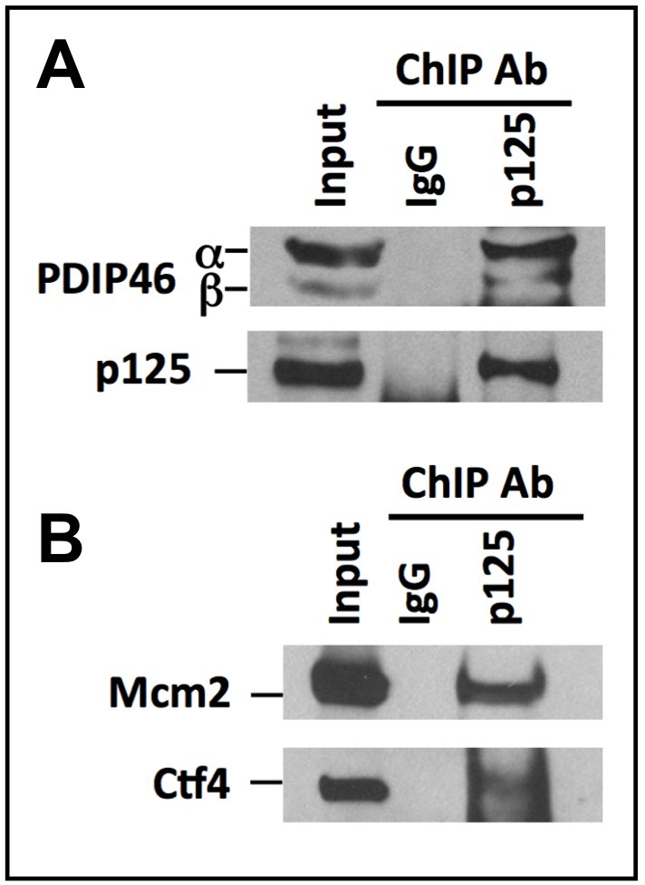
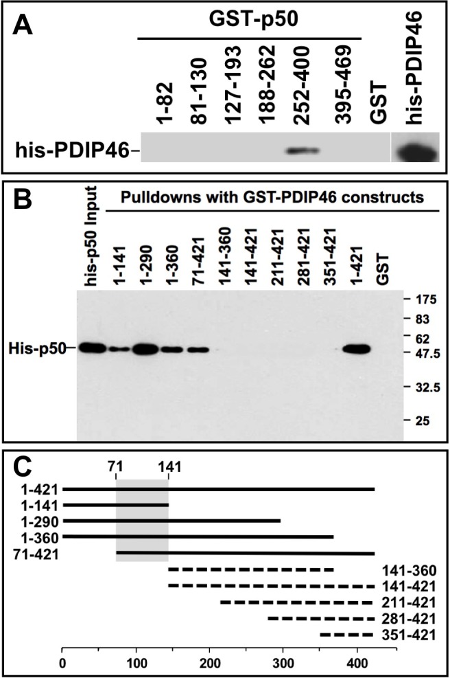
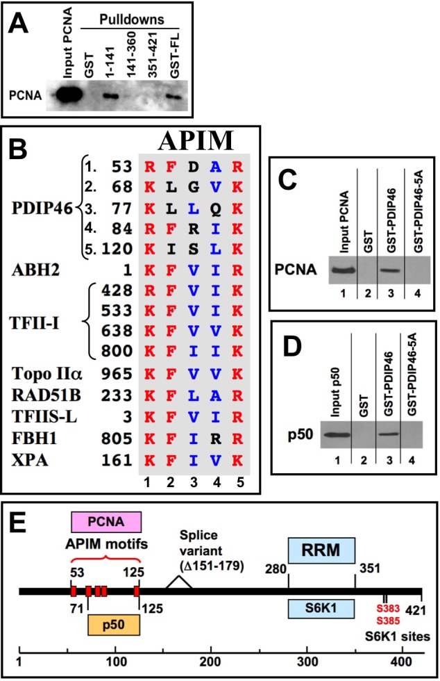
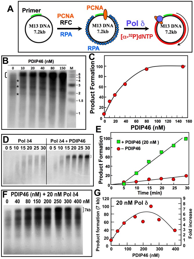
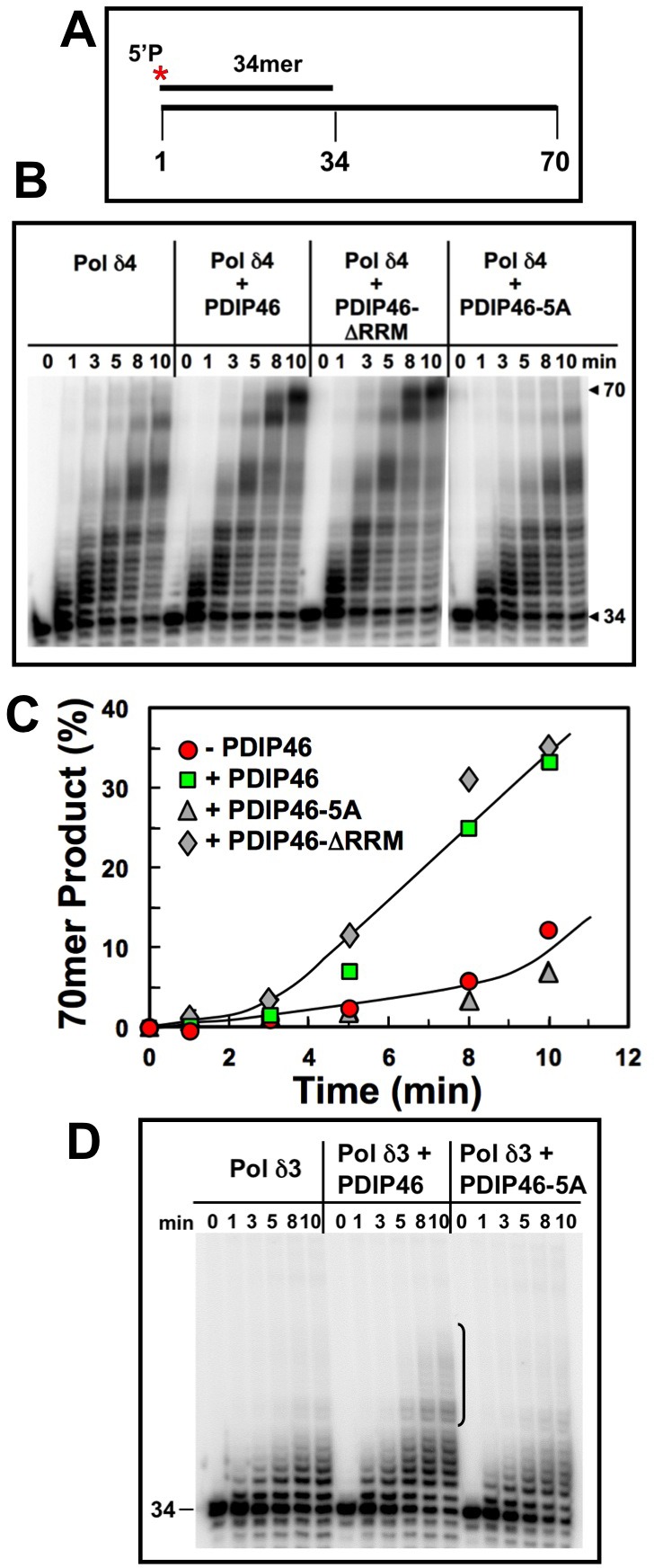
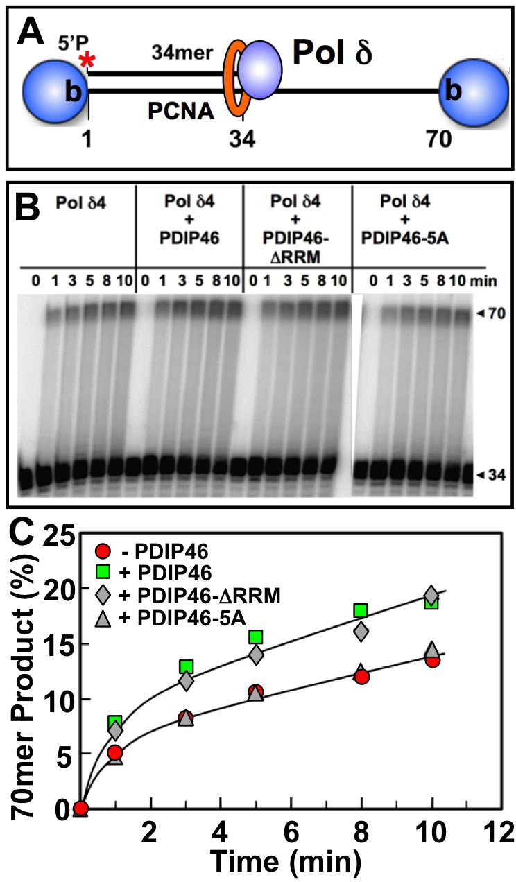
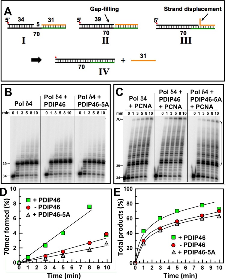
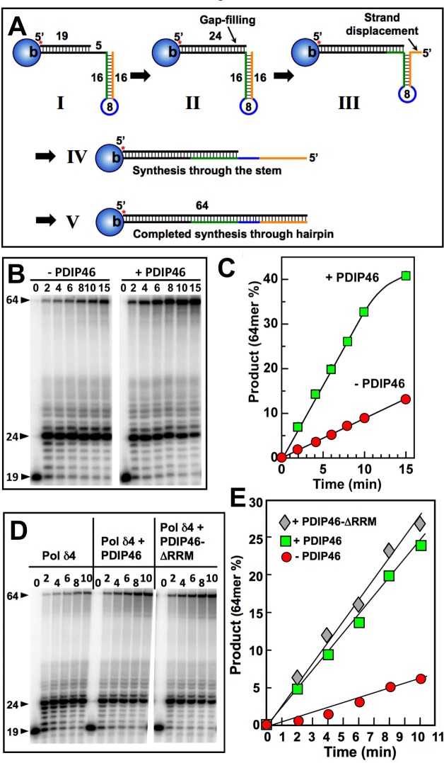
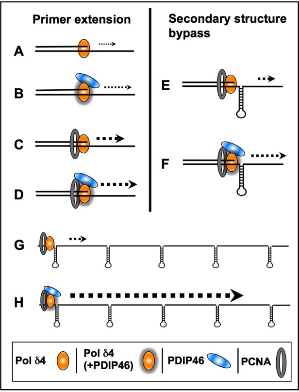
PDIP46 (SKAR, POLDIP3) was discovered through its interaction with the p50 subunit of human DNA polymerase δ (Pol δ). Its functions in DNA replication are unknown. PDIP46 associates with Pol δ in cell extracts both by immunochemical and protein separation methods, as well as by ChIP analyses. PDIP46 also interacts with PCNA via multiple copies of a novel PCNA binding motif, the APIMs (AlkB homologue-2 PCNA-Interacting Motif). Sites for both p50 and PCNA binding were mapped to the N-terminal region containing the APIMs. Functional assays for the effects of PDIP46 on Pol δ activity on singly primed ssM13 DNA templates revealed that it is a novel and potent activator of Pol δ. The effects of PDIP46 on Pol δ in primer extension, strand displacement and synthesis through simple hairpin structures reveal a mechanism where PDIP46 facilitates Pol δ4 synthesis through regions of secondary structure on complex templates. In addition, evidence was obtained that PDIP46 is also capable of exerting its effects by a direct interaction with Pol δ, independent of PCNA. Mutation of the Pol δ and PCNA binding region resulted in a loss of PDIP46 functions. These studies support the view that PDIP46 is a novel accessory protein for Pol δ that is involved in cellular DNA replication. This raises the possibility that altered expression of PDIP46 or its mutation may affect Pol δ functions in vivo, and thereby be a nexus for altered genomic stability.

Keywords: DNA polymerase δ; DNA replication; PDIP46; POLDIP3; SKAR.


Conflict of interest statement

CONFLICTS OF INTEREST

None

Figures

Figure 1
Figure 1. Association of PDIP46 with Pol δ by co-immunopreciptation, gel filtration, and native gradient gel electrophoresis
A. PDIP46 interacts with the Pol δ4 holoenzyme. GST-PDIP46 (GST-p46) was used to pull-down purified Pol δ. The pull-downs were western blotted for the p125, p68, p50, and p12 subunits of Pol δ. B. Co-immunoprecipitation of PDIP46 with Pol δ. HeLa cell extracts were immunoprecipitated with antibodies against PDIP46, and western blotted for the p50 (left panel) and p125 subunits (right panel) of Pol δ. C. Nuclear extracts of HEK 293 cells were chromatographed on a Superose 6 FPLC column as previously described [35]. Column fractions were western blotted for p125 and PDIP46. “BC” refers to the nuclear extract. Positions of molecular weight standards are shown on the left. The arrows refer to the elution of protein standards (Mr: thyroglobulin, 669,000; ferritin, 440,000; aldolase, 158,000). D. HEK 293 cell lysates were subjected to native (nondenaturing) gradient gel electrophoresis until limiting mobility was reached. Proteins were transferred to nitrocellulose membranes and Western blotted for p125 and PDIP46 (Materials and Methods). The migration positions of marker proteins (thyroglobulin, ferritin and catalase) and their respective native molecular weights are shown on the left.
Figure 2
Figure 2. PDIP46 is associated with chromatin bound Pol δ by ChIP analysis using antibody against p125
ChIP analysis was performed with A549 cells as described in “Materials and Methods”. A. The immunoprecipitates were western blotted with antibodies against PDIP46 and the p125 subunit of Pol δ. IgG refers to the control immunoprecipitation with non-immune serum. The bands marked “α” and “β” refer to the full-length PDIP46 and its minor spliced variant, respectively. B. ChIP analysis was performed as in (A) and western blotted for MCM2 and Ctf4 which were used as positive controls.
Figure 3
Figure 3. Mapping of the interaction sites between p50 and PDIP46
A. Mapping of the region of p50 that interacts with PDIP46. GST-fusion constructs of p50 were used to pull-down his-PDIP46, and western blotted for PDIP46. Only the deletion mutant containing residues 252-400 of p50 interacted with PDIP46. B. Mapping of the PDIP46 region that interacts with the p50 subunit of Pol δ. GST-PDIP46 deletions were used for pull-down assays of his-p50, and western blotted for p50. C. Diagrammatic summary of the data of panel B. Solid bars show those deletion constructs that interacted with p50, and the dashed lines show those that did not. The shaded area shows the common region of the PDIP46 deletion mutants that interacted with p50 (residues 71-141).
Figure 4
Figure 4. PDIP46 interacts with PCNA, and does so via APIM motifs
A. Mapping of the PDIP46 domain that interacts with PCNA. GST-PDIP46 deletion mutants were used to pull-down PCNA, and western blotted for PCNA. Only the GST-1-141 fusion protein interacted with PCNA. B. The N-terminus of PDIP46 harbors 5 APIM motifs. The alignment shows the five APIM motifs of PDIP46, together with those of the oxidative demethylase ABH2, the four APIMs of TFII-I, Topo IIα, Rad51B and TFIIS-L [34], FBH1 (F-box helicase) [44] and XPA [43]. Residues in red show the conserved basic residues at positions 1 and 5, as well as the phenylalanine at position 2, while those in blue are the aliphatic residues at positions 3 and 4. All sequences shown are those of human proteins. C. Mutation of the APIMs of PDIP46 leads to loss of PCNA binding. The conserved residues in positions 1, 2 and 5 of the APIM motifs were mutated to alanines (PDIP46-5A). GST-PDIP46 and the GST-PDIP46-5A mutant were used in pull-down assays of PCNA. D. Mutation of the APIMs of PDIP46 also leads to loss of p50 binding. GST-PDIP46 and the GST-PDIP46-5A mutant were used in pull-down assays of his-p50. E. Domain map of PDIP46 showing the location of binding regions (boxed) for p50, PCNA (APIM motifs), RRM, and S6K1. The two S6K1 phosphorylation sites and the region deleted in a minor splice variant [29] are also shown.
Figure 5
Figure 5. PDIP46 stimulates product formation by Pol δ4 in the M13 assay
A. Diagram of the M13 assay of Pol δ activity. Singly primed M13 ssDNA (left) is loaded with PCNA with RFC, and RPA single stranded DNA binding protein (center); Pol δ4 and [α-32P]-dATP is added to extend the primer up to the full-length product (right). B. Effects of increasing concentrations of PDIP46 on Pol δ4 activity. Pol δ4 concentration was 5 nM, M13 ssDNA was 2.5 nM, and PCNA was 6 nM (Materials and Methods). Reactions were incubated at 37° C for 25 min. Products were analyzed by electrophoresis on 1.2% alkaline agarose gels and were visualized by phosphorimaging. Lane M shows the migration of the markers. The bracket on the top left indicates the region that was used for quantitation of full-length 7 kb products. The asterisks show bands where pausing of the reactions occurred. C. Full-length product formation for panel B was quantified, and plotted as relative product formation against PDIP46 concentration. The data were fitted to a one site binding hyperbola using Prism software, and gave an apparent KD of 34 ± 7.7 nM (R2 = 0.98). D. Time dependence of product formation by Pol δ4 in the M13 assay in the presence of PDIP46. Pol δ4 (10 nM) was assayed on singly primed M13 in the absence (left panel) and presence (right panel) of PDIP46 (20 nM); the reactions were analyzed after 5, 10, 15, 20, 25 and 30 min. E. Product formation of the full-length products in panel D was quantified, and plotted as relative product formation against time. Data in the absence of PDIP46 are shown as circles, and those in the presence of 20 nM PDIP46 are shown as squares. F. The effects of higher concentrations (0-400 nM) of PDIP46 on Pol δ (20 nM) assayed on the M13 substrate. Reaction times were 15 min. G. The full-length products for panel F were quantified and plotted against PDIP46 concentration.
Figure 6
Figure 6. PDIP46 and PDIP46-ΔRRM but not PDIP46-5A stimulate primer extension by Pol δ4 on oligonucleotide substrates in the absence of PCNA
A. Oligonucleotide substrate for primer extension. A 5′-[32P]end-labeled 34mer primer was annealed to a 70mer template. The asterisk denotes the labeling. B. Effects of PDIP46 and PDIP46-5A (50 nM) on primer extension by Pol δ4 in the absence of PCNA. The concentration of reactants were DNA substrate (100 nM), Pol δ4 or Pol δ3 (5 nM), PDIP46, PDIP46-ΔRRM or PDIP46-5A (50 nM). The reactions were performed for times ranging from 0-10 min. Reaction products were resolved by electrophoresis on sequencing gels and visualized by phosphorimaging (Materials and Methods.). C. The full-length 70mer primer extension products for panel B were quantified and plotted (as % of primer converted to 70mer) against time. Data for Pol δ4 in the absence of PDIP46 is shown as solid circles, with PDIP46 as solid squares, with PDIP46-ΔRRM as shaded diamonds and with PDIP46-5A as shaded triangles. D. Effects of PDIP46 and PDIP46-5A on Pol δ3 activity in the absence of PCNA. Reactions were performed as described in B. The vertical bracket on the gel show a region of primer extension that is increased in the presence of PDIP46.
Figure 7
Figure 7. Effects of PDIP46, PDIP46-ΔRRM and PDIP46-5A on primer extension by Pol δ on oligonucleotide substrates in the presence of PCNA
A. Oligonucleotide substrate for primer extension. A 5′-[32P]end-labeled 34mer primer was annealed to a 70mer template, and was then blocked with streptavidin (shaded sphere). PCNA was then loaded onto the substrate with RFC. B. Effects of PDIP46 and PDIP46-5A (50 nM) on primer extension by Pol δ4 in the presence of PCNA. The concentrations of reactants were DNA (50 nM), Pol δ4 (10 nM), PCNA (50 nM), PDIP46 or its mutants (50 nM). Conditions used were as described in Materials and Methods. The reactions were performed for times ranging from 0-10 min. Reaction products were resolved by electrophoresis on sequencing gels and visualized by phosphorimaging. C. Amounts of 70mer formed in panel B were determined and plotted against time. Data for Pol δ4 in the absence of PDIP46 is shown as solid circles, with PDIP46 as solid squares, with PDIP46-ΔRRM as shaded diamonds and with the PDIP46-5A mutant as shaded triangles.
Figure 8
Figure 8. PDIP46 stimulates strand displacement by Pol δ4
A. Oligonucleotide substrate for primer strand displacement assays. A 5′-[32P]end labeled 34mer primer was annealed to a 70mer template as in Figure 6, together with a downstream blocking 31mer to leave a 5nt gap. The asterisk denotes the labeling. The concentration of reactants were DNA template (100 nM), Pol δ4 (5 nM), PDIP46 or PDIP46-5A (50 nM), and PCNA (100 nM) when added. Reactions were performed for the indicated times. B. Effects of PDIP46 and PDIP46-5A (50 nM) on strand displacement by Pol δ4 in the absence of PCNA. Reaction products were visualized by phosphorimaging. C. Effects of PDIP46 and PDIP46-5A on strand displacement by Pol δ4 in the presence of PCNA. Reaction products were visualized by phosphorimaging. D. The 70mer full-length primer extension products for C, reflecting complete strand displacement of the blocking 31mer oligonucleotide, were quantified and plotted as 70mer formed as % of primer against time. Data points in the absence of PDIP46 are shown as solid circles, with PDIP46 as solid squares, and with PDIP46-5A as shaded triangles. E. The overall strand displacement products as reflected by primer extension products from the 40mer-70mer (indicated by the bracket in C) were quantified and plotted against time. Data points were labeled as for panel D.
Figure 9
Figure 9. PDIP46 stimulates primer extension by Pol δ4 through a template with a hairpin
A Diagram of the substrate and expected progression through the stem loop structure. For details see text. B. Pol δ4 (15 nM) was reacted for the indicated times with the substrate (50 nM) in the absence and presence of PDIP46 (50 nM) after the loading of PCNA with RFC (Experimental Procedures). The products were analyzed by 8M urea denaturing polyacrylamide gel electrophoresis and visualized by phosphorimaging. The arrowheads indicate the positions of the primer (19nt), the position at the point of primer extension to the 5′-end of the template at the start of the hairpin (24nt), and the full-length product (64nt). C. The amounts of 64mer representing synthesis through the hairpin for the phosphorimage in B were quantified. Data are plotted as percentage of primer converted in the absence (solid circles) and presence (solid squares) of PDIP46. D. The RRM region is not required for PDIP46 stimulation of Pol δ4. The effects of PDIP46 and the PDIP46-ΔRRM mutant were examined. The concentrations of the reactants were DNA (50 nM), Pol δ4 (10 nM), and PDIP46 or PDIP46-ΔRRM (50 nM). E. Formation of the 64mer for panel D was quantified and plotted against time. Data for the control are shown as solid circles, for PDIP46 as solid squares, and for PDIP46-ΔRRM as solid diamonds.
Figure 10
Figure 10. Diagrammatic summary of the effects of PDIP46 on Pol δ4 activity
(A., B.) PDIP46 stimulates Pol δ4 in the absence of PCNA, revealing a direct effect on Pol δ4. (C., D.) Pol δ4 synthesis in the presence of PCNA. Pol δ4 is strongly stimulated by PCNA alone, due to its conversion to a processive mode of synthesis, but this is further stimulated by PDIP46. (E., F.) PDIP46 stimulates PCNA-enabled synthesis through a hairpin secondary structure. (This is similar to effects in strand displacement, which are omitted). It is proposed that there is an additive effect of PDIP46 on Pol δ4 synthesis over that observed with oligonucleotides with a single secondary structure; this is illustrated in G. and H., to show the additive nature of the facilitation of synthesis when multiple stem-loop/hairpin structures are present. This provides a working hypothesis for the potent stimulation of Pol δ4 synthesis of full-length products in the M13 assay by PDIP46. The increases in product formation are qualitatively represented by increases in weight of the dotted arrows for all panels. The direct effects of PDIP46 on Pol δ that indicate alteration in Pol δ function are shown by shadowing of the icons for Pol δ4 (B, D, F).

Similar articles

Cited by

References

    1. Kunkel TA, Bebenek K. DNA replication fidelity. Annu Rev Biochem. 2000;69:497–529. - PubMed
    1. Loeb LA, Monnat RJJ. DNA polymerases and human disease. Nat Rev Genet. 2008;9:594–604. - PubMed
    1. Prindle MJ, Loeb LA. DNA polymerase delta in DNA replication and genome maintenance. Environ Mol Mutagen. 2012;53:666–682. - PMC - PubMed
    1. Reha-Krantz LJ. DNA polymerase proofreading: Multiple roles maintain genome stability. Biochim Biophys Acta. 2010;1804:1049–1063. - PubMed
    1. Briggs S, Tomlinson I. Germline and somatic polymerase epsilon and delta mutations define a new class of hypermutated colorectal and endometrial cancers. J Pathol. 2013;230:148–153. - PMC - PubMed

Publication types