. 2016 Mar 18;44(5):2393-408.
doi: 10.1093/nar/gkw023. Epub 2016 Jan 26.

Novel RNA-binding activity of MYF5 enhances Ccnd1/Cyclin D1 mRNA translation during myogenesis

Affiliations

Novel RNA-binding activity of MYF5 enhances Ccnd1/Cyclin D1 mRNA translation during myogenesis

Amaresh C Panda et al. Nucleic Acids Res. .

Abstract

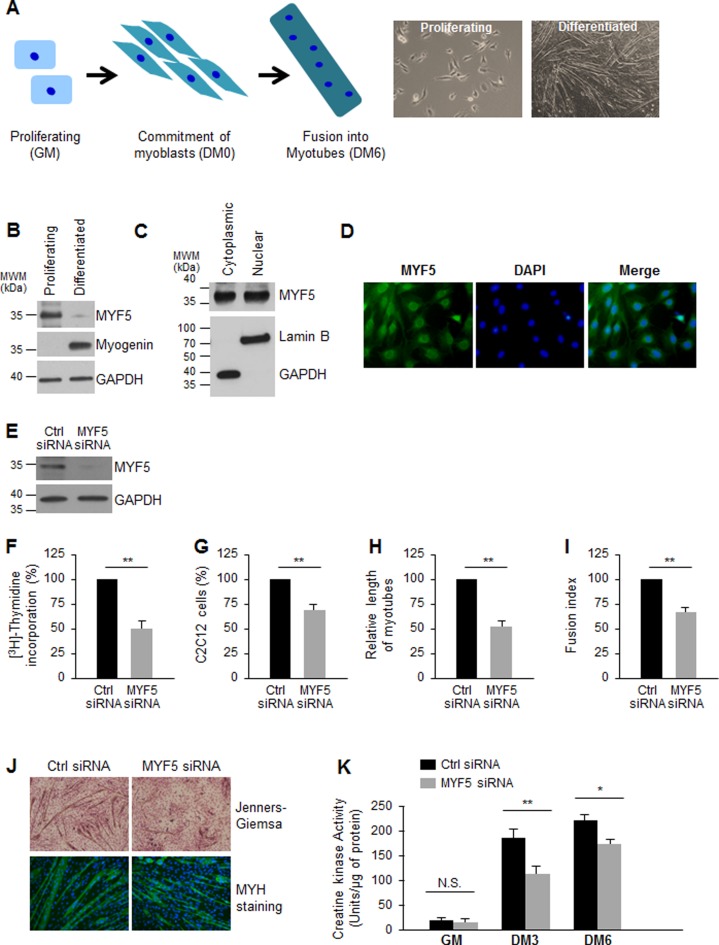
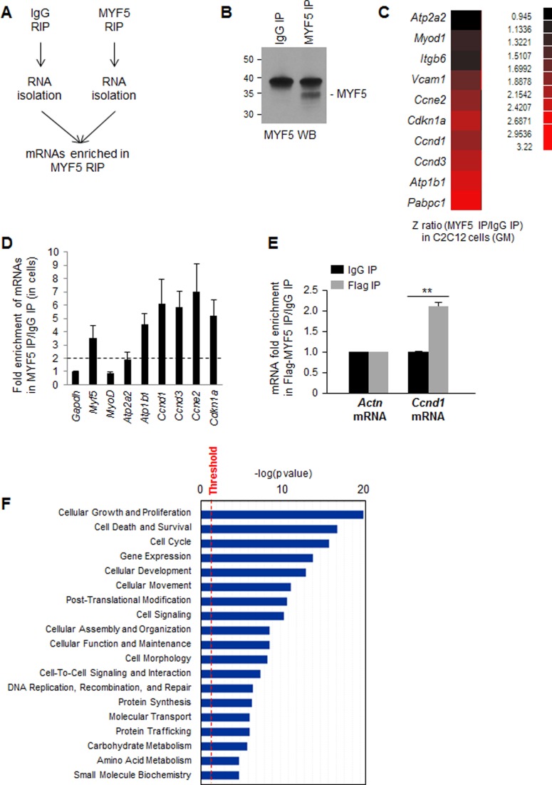
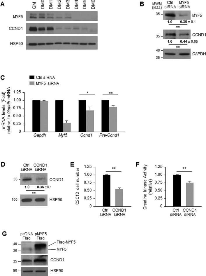
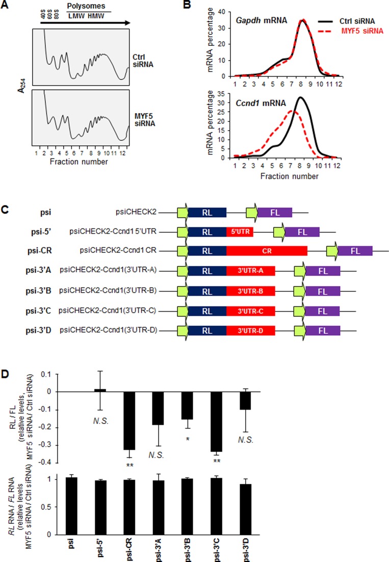
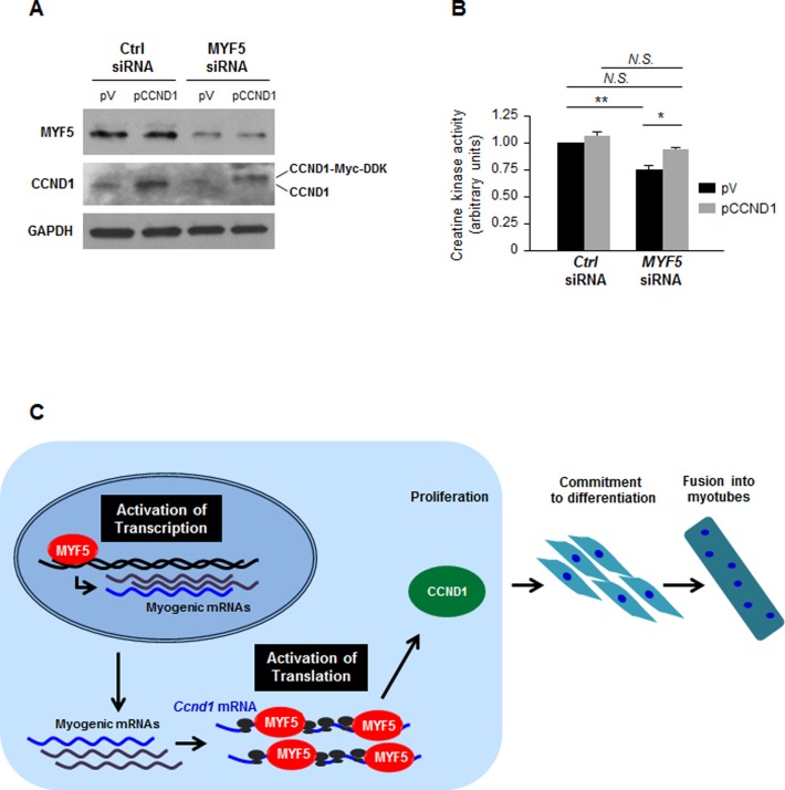
Skeletal muscle contains long multinucleated and contractile structures known as muscle fibers, which arise from the fusion of myoblasts into multinucleated myotubes during myogenesis. The myogenic regulatory factor (MRF) MYF5 is the earliest to be expressed during myogenesis and functions as a transcription factor in muscle progenitor cells (satellite cells) and myocytes. In mouse C2C12 myocytes, MYF5 is implicated in the initial steps of myoblast differentiation into myotubes. Here, using ribonucleoprotein immunoprecipitation (RIP) analysis, we discovered a novel function for MYF5 as an RNA-binding protein which associated with a subset of myoblast mRNAs. One prominent MYF5 target was Ccnd1 mRNA, which encodes the key cell cycle regulator CCND1 (Cyclin D1). Biotin-RNA pulldown, UV-crosslinking and gel shift experiments indicated that MYF5 was capable of binding the 3' untranslated region (UTR) and the coding region (CR) of Ccnd1 mRNA. Silencing MYF5 expression in proliferating myoblasts revealed that MYF5 promoted CCND1 translation and modestly increased transcription of Ccnd1 mRNA. Accordingly, overexpressing MYF5 in C2C12 cells upregulated CCND1 expression while silencing MYF5 reduced myoblast proliferation as well as differentiation of myoblasts into myotubes. Moreover, MYF5 silencing reduced myogenesis, while ectopically restoring CCND1 abundance partially rescued the decrease in myogenesis seen after MYF5 silencing. We propose that MYF5 enhances early myogenesis in part by coordinately elevating Ccnd1 transcription and Ccnd1 mRNA translation.


Figures

Figure 1.
Figure 1.
MYF5 is necessary for C2C12 myoblast growth and differentiation: (A)Left, Schematic of skeletal muscle differentiation. Proliferating myoblasts become committed to differentiation, exit the cell division cycle and begin to elongate, subsequently fusing together to form multinucleated myotubes. Right, phase-contrast images of proliferating C2C12 myoblasts cultured in growth medium (GM) and C2C12 cultures displaying myotubes after culture in differentiation medium [(DM) DMEM with 2% horse serum] for 6 days. (B) Western blot analysis of the levels of MYF5 (and loading control GAPDH) in proliferating and differentiated C2C12 myoblasts. (C) Western blot analysis of MYF5 localization in proliferating C2C12 myoblasts; GAPDH and Lamin B were assessed as cytoplasmic and nuclear fractionation markers, respectively. (D) Immunofluorescence detection of MYF5 in growing C2C12 myoblasts; nuclei were visualized by staining with DAPI. (E) Western blot analysis of MYF5 and GAPDH expression levels 48 h after transfection of proliferating C2C12 myoblasts with Ctrl siRNA or MYF5 siRNA. (F,G) Forty-eight hours after transfection of C2C12 myoblasts with Ctrl siRNA or MYF5 siRNA, measurements were taken for [3H]-thymidine incorporation (F) and cell numbers (using a TC10 automated cell counter, BioRad) (G). (H,I) After differentiation of C2C12 cells in which MYF5 levels were normal (Ctrl siRNA) or was reduced (MYF5 siRNA) through DM6, the length of myotubes (H) and the fusion index (fraction of total nuclei present myotube) (I) were assessed. (J) Micrographs of Jenners-Giemsa stain (a dye that stains myotubes and nuclei) (top) and fluorescence micrographs to detect MYH (present in differentiated myotubes) in C2C12 cells that had been transfected with MYF5 siRNA or Ctrl siRNA and placed in differentiation medium for 6 days (DM6). (K) C2C12 differentiation was monitored using creatine kinase activity at different time points of differentiation: cells in proliferation (growth) medium (GM), and cells in differentiation medium for 3 or 6 days (DM3 and DM6). In (F-I,K), data are the means and S.E.M. from three independent experiments. N.S., not significant; *, P < 0.05; **, P < 0.01 (Student's t-test).
Figure 2.
Figure 2.
MYF5 target transcripts include mRNAs that encode proteins involved in myoblast proliferation and differentiation. (A) RIP assay using cytoplasmic lysates prepared from C2C12 cells using either anti-MYF5 antibody or IgG under conditions that preserved mRNA-RBP (mRNP) complexes. (B) Western blot analysis of MYF5 recovered in IP samples. (C) Following MYF5 RIP, MYF5-bound mRNAs were identified by microarray (RIP-chip) analysis in growing (GM) C2C12 cells. Data represent the Z-ratio of mRNAs in MYF5 RIP relative to IgG RIP. (D) RIP followed by RT-qPCR analysis to validate the association of MYF5 with mRNAs encoding myogenic proteins in proliferating C2C12 myoblasts; the levels of mRNAs in MYF5 IP were normalized to the levels of Gapdh mRNA and plotted as fold enrichment relative to the levels seen in control IgG IP samples. Discontinuous gray line: twofold enrichment in mRNAs bound to MYF5. (E) RIP analysis of Flag-MYF5 interaction with Ccnd1 mRNA. Forty-eight hours after C2C12 transfection with Flag-MYF5, RIP analysis was carried out using IgG or anti-Flag antibodies. Ccnd1 mRNA was detected by RT-qPCR analysis and its levels in Flag IP were compared with those in control IgG IP samples; Actn mRNA (encoding the housekeeping protein β-Actin) was measured to normalize sample input. Data in (D,E) represent the means and S.E.M. from three or more independent experiments. (F) Ingenuity pathway analysis (IPA) of mRNAs enriched in MYF5 IP relative to IgG in C2C12 growing myoblasts. *, P < 0.05 and **, P < 0.01 (Student's t-test).
Figure 3.
Figure 3.
MYF5 binds to specific sequences on the Ccnd1 mRNA. (A)Top, schematic showing biotinylated RNA fragments spanning the 5′UTR, coding region (CR), and 3′UTR of Ccnd1 mRNA used for pulldown. Bottom, biotinylated RNA fragments were incubated with cytoplasmic lysates from C2C12 cells (GM); after pulldown using streptavidin beads, the levels of MYF5 bound to the biotinylated RNA segments were detected by western blot analysis. (B) Recombinant purified His-MYF5 was incubated with biotinylated Ccnd1 RNA fragments followed by pulldown and detection of MYF5 by western blot analysis using anti-MYF5 antibody. (C) Schematic of biotinylated RNA fragments spanning the Ccnd1 3′UTR-C transcript (top), were tested for binding to His-MYF5 after pull-down using streptavidin beads; His-MYF5 interaction with RNA segments of fragment C (middle) and smaller RNAs after closer subdivision of fragments 9 through 11 (bottom) were assessed by western blot analysis using anti-MYF5 antibody. (D) GST or GST-MYF5 were incubated with radiolabeled Ccnd1 3′UTR-C10–1, then either resolved on native acrylamide gels (left) by RNA electrophoretic mobility shift assay (EMSA), or crosslinked by UV irradiation and resolved by SDS-PAGE (right). (E) The domain of MYF5 that interacts with the Ccnd1 3′-C fragment was mapped by creating GST-tagged truncations of MYF5 (left) and testing their interaction by biotin pulldown and western blot analysis using anti-GST antibody (right).
Figure 4.
Figure 4.
MYF5 regulates CCND1 expression in C2C12 myoblasts. (A) Western blot analysis of MYF5 and CCND1 expression during C2C12 differentiation; heat shock protein 90 (HSP90) was included as loading control. (B,C) Forty-eight hours after transfecting C2C12 cells with MYF5 siRNA or Ctrl siRNA, the levels of MYF5, CCND1 and loading control GAPDH were analyzed by western blot analysis (B) and the levels of Ccnd1 pre-mRNA and mRNA by RT-qPCR analysis (C). (D–F) Forty-eight hours after transfection of proliferating C2C12 myoblasts with Ctrl or CCND1 siRNAs, the levels of CCND1 and HSP90 were assessed by western blot analysis (D) and cell numbers were measured using a TC10 automated cell counter (BioRad) and represented as fold change in cell number after CCND1 silencing relative to those in the Ctrl siRNA group (E). At day 6 in differentiation medium (DM6) C2C12 differentiation was monitored by measuring creatine kinase activity (F). (G) Forty-eight hours after MYF5 was overexpressed using pFlag-MYF5 pcDNA3, the levels of MYF5, CCND1 and loading control HSP90 were studied by western blot analysis. Data in (B-F) represent the means and S.E.M. from three or four independent experiments. *, P < 0.05 and **, P < 0.01 (Student's t-test).
Figure 5.
Figure 5.
MYF5 promotes translation of Ccnd1 mRNA. (A,B) Forty-eight hours after Ctrl or MYF5 siRNA transfection of C2C12 cells, cytoplasmic extracts were fractionated through sucrose gradients to obtain cytoplasmic components of progressively larger weight: ribosomal subunits (40S, 60S), monosomes (80S) and low-molecular-weight (LMW) and high-molecular-weight (HMW) polysomes (A). The relative distribution of Gapdh mRNA, encoding a housekeeping protein, and Ccnd1 mRNA were measured by RT-qPCR analysis of RNA in each of the gradient fractions and represented as percentage of total RNA in the gradient (B). (C) Schematic of the dual luciferase reporter plasmids derived from the parent vector psiCHECK2 (psi), which expresses renilla luciferase (RL) and the internal control firefly luciferase (FL), and psiCHECK2-derived plasmids bearing the Ccnd1 fragments downstream of the RL coding region. (D)Top, Influence of MYF5 silencing on the expression of the reporter constructs. Twenty-four h after transfection of C2C12 cells with either MYF5 siRNA or Ctrl siRNA, each reporter plasmid was transfected, and 16 h later the ratio of RL activity to FL activity was measured. The decrease in relative RL/FL ratio of MYF5 siRNA-transfected cells relative to the RL/FL ratio of Ctrl siRNA-transfected cells is indicated. Bottom, RT-qPCR analysis of RL mRNA levels normalized to FL mRNA levels in each transfection group. Data represent the means and S.E.M. from 3 independent experiments. N.S., not significant; *, P < 0.05; **, P < 0.01 (Student's t-test).
Figure 6.
Figure 6.
Influence of MYF5 on myogenesis via regulation of CCND1 expression. (A,B) C2C12 cells were transfected with MYF5 siRNA or Ctrl siRNA, along with a control vector [pcDNA3-Flag (pV)] or a plasmid vector that expressed Myc-tagged CCND1. Forty-eight hours later, the levels of CCND1 and MYF5 were analyzed by western blot analysis (A) and the degree of differentiation was analyzed by measuring creatine kinase activity at day 6 into differentiation (B). Data presented are the means and S.E.M. from four independent experiments; significance (P) is indicated. (C) Proposed model whereby MYF5 modulates myogenesis by acting upon CCND1 expression on two levels: first, MYF5 activates Ccnd1 transcription moderately, and second, MYF5 binds the Ccnd1 mRNA at the CR and 3′UTR, promoting Ccnd1 mRNA translation. The net effect is a CCND1-mediated increase in myoblast proliferation necessary at the initiation of myogenesis. N.S., not significant; *, P < 0.05; **, P < 0.01 (Student's t-test).

References

    1. Collins C.A., Olsen I., Zammit P.S., Heslop L., Petrie A., Partridge T.A., Morgan J.E. Stem cell function, self-renewal, and behavioral heterogeneity of cells from the adult muscle satellite cell niche. Cell. 2005;122:289–301. - PubMed
    1. Moss F.P., Leblond C.P. Satellite cells as the source of nuclei in muscles of growing rats. Anat. Rec. 1971;170:421–435. - PubMed
    1. Tedesco F.S., Dellavalle A., Diaz-Manera J., Messina G., Cossu G. Repairing skeletal muscle: regenerative potential of skeletal muscle stem cells. J. Clin. Invest. 2010;120:11–19. - PMC - PubMed
    1. Chargé S.B., Rudnicki M.A. Cellular and molecular regulation of muscle regeneration. Physiol. Rev. 2004;84:209–238. - PubMed
    1. Hawke T.J., Garry D.J. Myogenic satellite cells: physiology to molecular biology. J. Appl. Physiol. 2001;91:534–551. - PubMed

Publication types

MeSH terms