

Early treatment of HER2-amplified brain tumors with targeted NK-92 cells and focused ultrasound improves survival
- PMID: 26819443
- PMCID: PMC4896543
- DOI: 10.1093/neuonc/nov318
Early treatment of HER2-amplified brain tumors with targeted NK-92 cells and focused ultrasound improves survival
Abstract
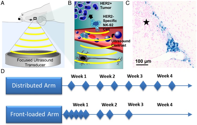
Background: Malignant brain tumors have a dismal prognosis, with residual tumor remaining after surgery necessitating adjuvant chemoradiotherapy. The blood-brain barrier hinders many chemotherapeutic agents, resulting in modest treatment efficacy. We previously demonstrated that targeted natural killer (NK)-92 cells could be delivered to desired regions of the brain using MRI-guided focused ultrasound and Definity microbubbles. Targeted NK-92 cells have advantages over many systemic therapies including their specific cytotoxicity to malignant cells (particularly those expressing the target antigen), ability to spare healthy cells, and being unaffected by efflux channels.
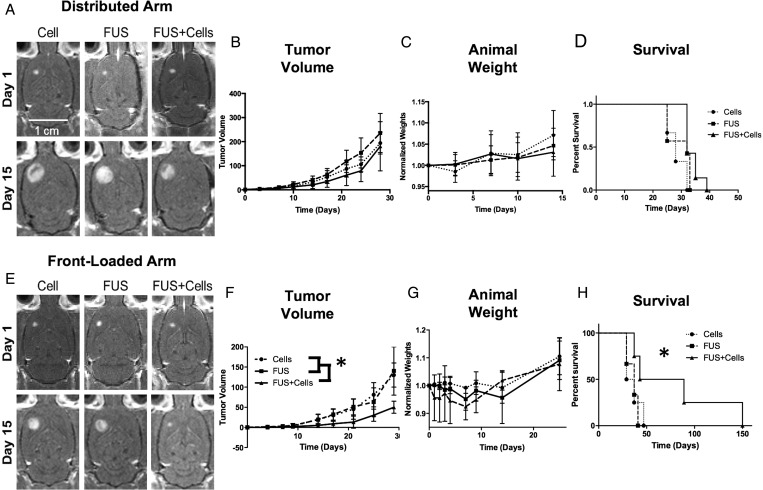
Methods: We investigated whether longitudinal treatments with targeted NK-92 cells, focused ultrasound, and microbubbles could slow tumor growth and improve survival in an orthotopic HER2-amplified rodent brain tumor model using a human breast cancer line as a prototype. The HER2 receptor, involved in cell growth and differentiation, is expressed by both primary and metastatic brain tumors. Breast cancers with HER2 amplification have a higher risk of CNS metastasis and poorer prognosis.
Results: Early intensive treatment with targeted NK-92 cells and ultrasound improved survival compared with biweekly treatments or either treatment alone. The intensive treatment paradigm resulted in long-term survival in 50% of subjects.
Conclusions: Many tumor proteins could be exploited for targeted therapy with the NK-92 cell line; combined with the mounting safety evidence for transcranial ultrasound, these results may soon be translatable to a highly targeted treatment option for patients with brain tumors.
Keywords: MRIgFUS; breast cancer; focused ultrasound; immune cell therapy.
© The Author(s) 2016. Published by Oxford University Press on behalf of the Society for Neuro-Oncology. All rights reserved. For permissions, please e-mail: journals.permissions@oup.com.
Figures
Similar articles
-
Ultrasound-mediated blood-brain/blood-tumor barrier disruption improves outcomes with trastuzumab in a breast cancer brain metastasis model.J Control Release. 2012 Nov 10;163(3):277-84. doi: 10.1016/j.jconrel.2012.09.007. Epub 2012 Sep 18. J Control Release. 2012. PMID: 23000189 Free PMC article.
-
Focused ultrasound delivers targeted immune cells to metastatic brain tumors.Cancer Res. 2013 Mar 15;73(6):1892-9. doi: 10.1158/0008-5472.CAN-12-2609. Epub 2013 Jan 9. Cancer Res. 2013. PMID: 23302230 Free PMC article.
-
Growth inhibition in a brain metastasis model by antibody delivery using focused ultrasound-mediated blood-brain barrier disruption.J Control Release. 2016 Sep 28;238:281-288. doi: 10.1016/j.jconrel.2016.08.001. Epub 2016 Aug 3. J Control Release. 2016. PMID: 27496633 Free PMC article.
-
A review of potential applications of MR-guided focused ultrasound for targeting brain tumor therapy.Neurosurg Focus. 2018 Feb;44(2):E10. doi: 10.3171/2017.11.FOCUS17620. Neurosurg Focus. 2018. PMID: 29385922 Review.
-
Ultrasound-induced blood-brain barrier disruption for the treatment of gliomas and other primary CNS tumors.Cancer Lett. 2020 Jun 1;479:13-22. doi: 10.1016/j.canlet.2020.02.013. Epub 2020 Feb 27. Cancer Lett. 2020. PMID: 32112904 Review.
Cited by
-
Chimeric antigen receptor natural killer cells: a promising antitumor immunotherapy.MedComm (2020). 2023 Dec 1;4(6):e422. doi: 10.1002/mco2.422. eCollection 2023 Dec. MedComm (2020). 2023. PMID: 38045827 Free PMC article. Review.
-
Focused ultrasound treatment for central nervous system disease: neurosurgeon's perspectives.Biomed Eng Lett. 2017 Jan 25;7(2):107-114. doi: 10.1007/s13534-017-0013-8. eCollection 2017 May. Biomed Eng Lett. 2017. PMID: 30603157 Free PMC article. Review.
-
Analysis of Multifrequency and Phase Keying Strategies for Focusing Ultrasound to the Human Vertebral Canal.IEEE Trans Ultrason Ferroelectr Freq Control. 2018 Dec;65(12):2322-2331. doi: 10.1109/TUFFC.2018.2872171. Epub 2018 Sep 26. IEEE Trans Ultrason Ferroelectr Freq Control. 2018. PMID: 30273151 Free PMC article.
-
Natural Killer Cells and Current Applications of Chimeric Antigen Receptor-Modified NK-92 Cells in Tumor Immunotherapy.Int J Mol Sci. 2019 Jan 14;20(2):317. doi: 10.3390/ijms20020317. Int J Mol Sci. 2019. PMID: 30646574 Free PMC article. Review.
-
Sonazoid-Conjugated Natural Killer Cells for Tumor Therapy and Real-Time Visualization by Ultrasound Imaging.Pharmaceutics. 2021 Oct 15;13(10):1689. doi: 10.3390/pharmaceutics13101689. Pharmaceutics. 2021. PMID: 34683982 Free PMC article.
References
-
- Nussbaum ES, Djalilian HR, Cho KH, Hall WA. Brain metastases. Histology, multiplicity, surgery, and survival. Cancer. 1996;78(8):1781–1788. - PubMed
-
- Hall WA, Djalilian HR, Nussbaum ES, Cho KH. Long-term survival with metastatic cancer to the brain. Med Oncol. 2000;17(4):279–286. - PubMed
-
- Ohgaki H, Dessen P, Jourde B et al. . Genetic pathways to glioblastoma: a population-based study. Cancer Res. 2004;64(19):6892–6899. - PubMed
-
- Stupp R, Mason WP, van den Bent MJ et al. . Radiotherapy plus concomitant and adjuvant temozolomide for glioblastoma. N Engl J Med. 2005;352(10):987–996. - PubMed
-
- Miralbell R, Mornex F, Greiner R et al. . Accelerated radiotherapy, carbogen, and nicotinamide in glioblastoma multiforme: report of European Organization for Research and Treatment of Cancer trial 22933. J Clin Oncol. 1999;17(10):3143–3149. - PubMed
Publication types
MeSH terms
Substances
Grants and funding
LinkOut - more resources
Full Text Sources
Other Literature Sources
Medical
Research Materials
Miscellaneous
