. 2016:2016:2632368.
doi: 10.1155/2016/2632368. Epub 2015 Dec 27.

Surgical Anatomy of the Gastrointestinal Tract and Its Vasculature in the Laboratory Rat

Affiliations

Surgical Anatomy of the Gastrointestinal Tract and Its Vasculature in the Laboratory Rat

Katarína Vdoviaková et al. Gastroenterol Res Pract. 2016.

Abstract

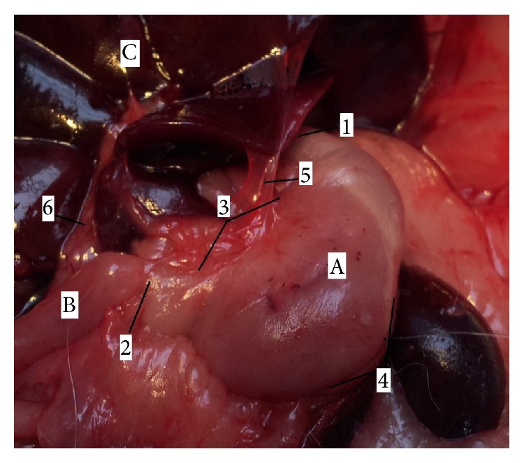
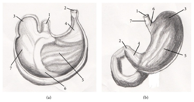
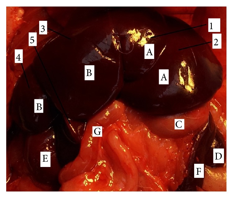
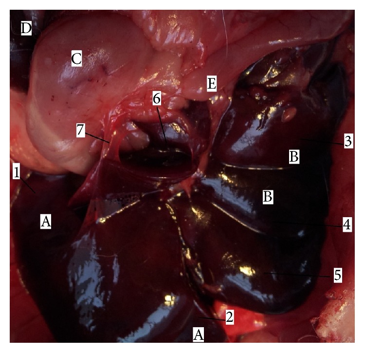
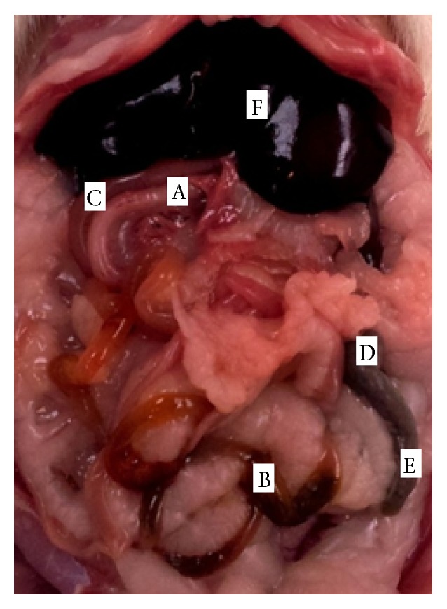
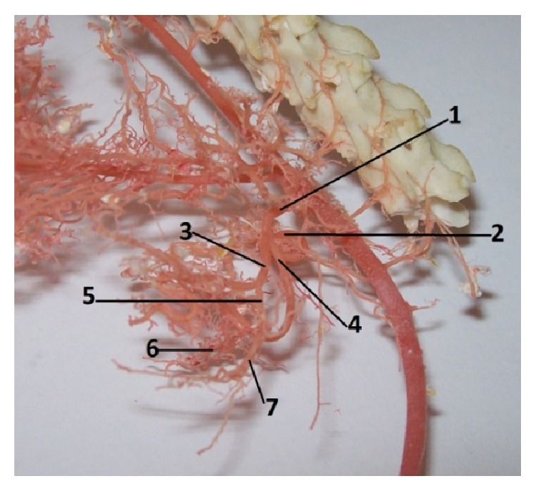
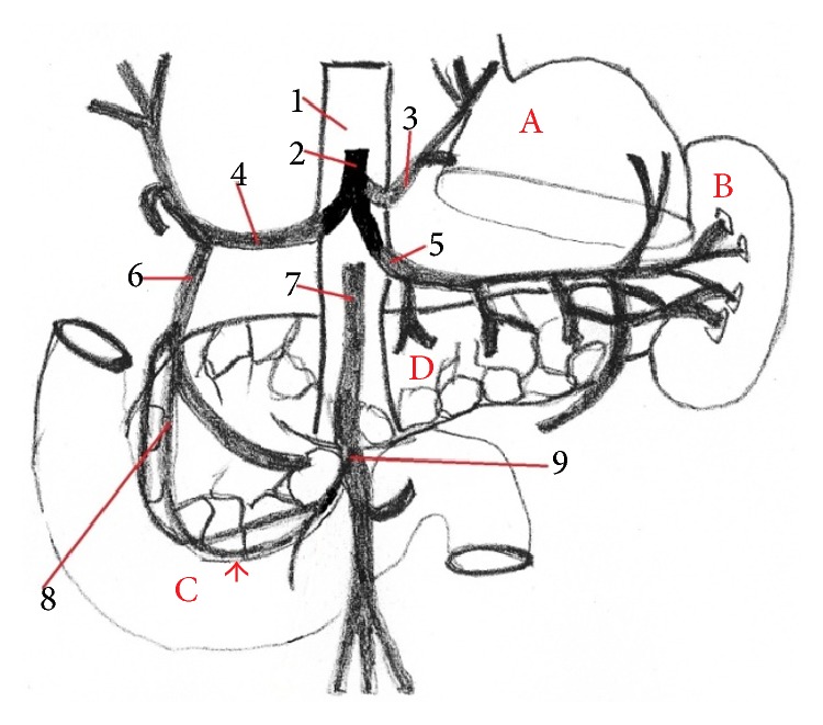
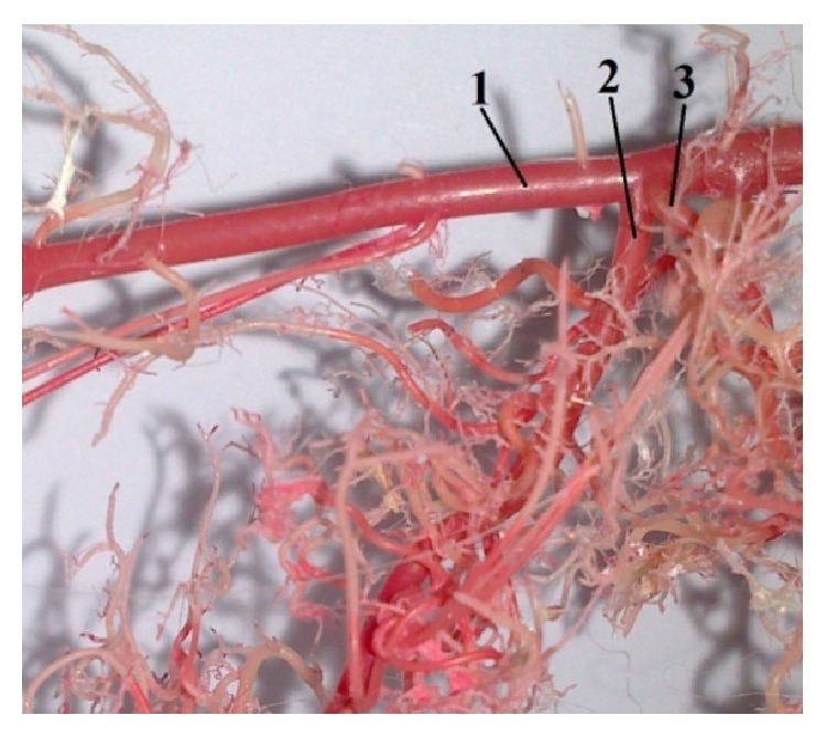
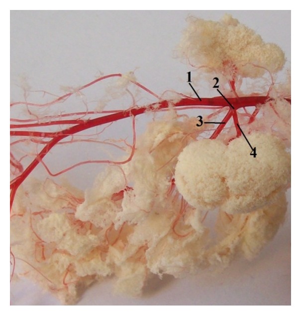
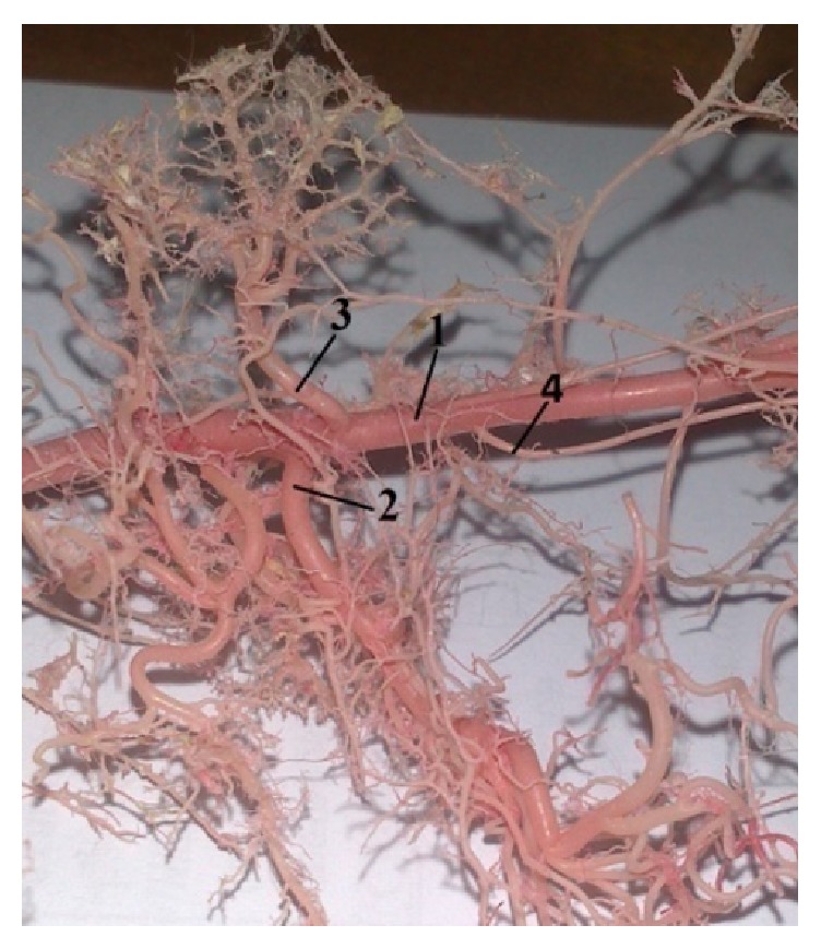
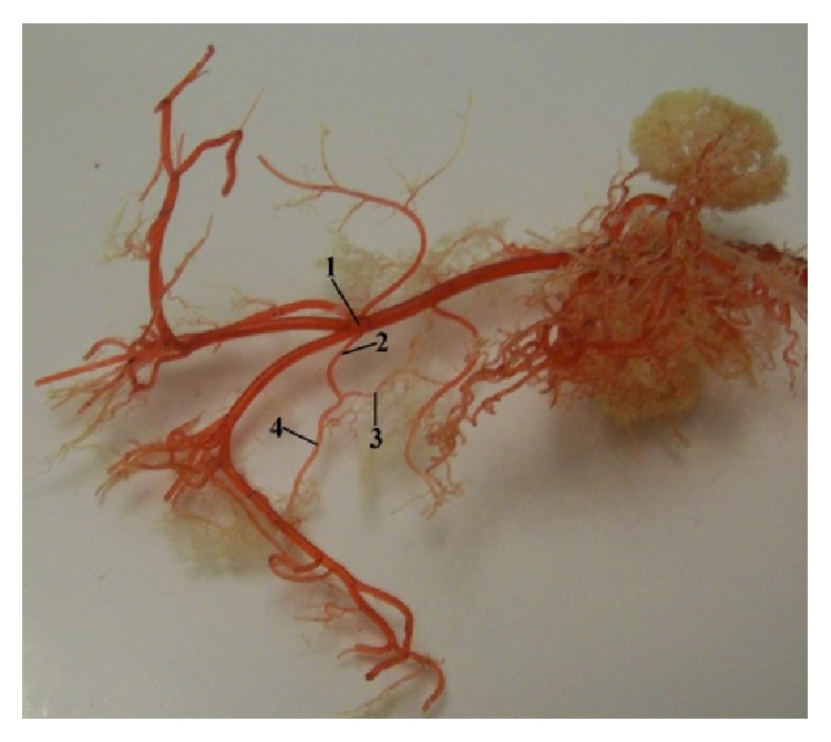
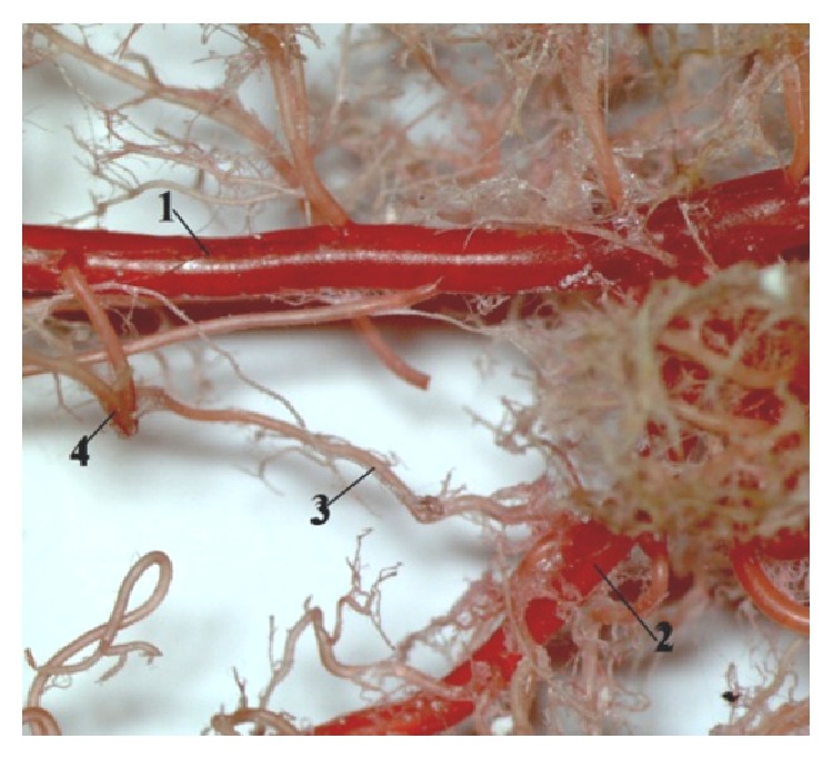
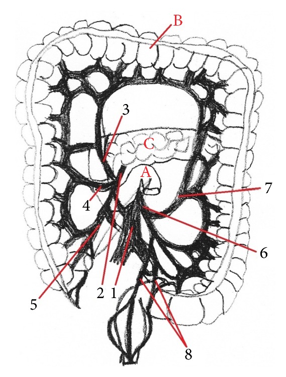
The aim of this study was to describe and illustrate the morphology of the stomach, liver, intestine, and their vasculature to support the planning of surgical therapeutic methods in abdominal cavity. On adult Wistar rats corrosion casts were prepared from the arterial system and Duracryl Dental and PUR SP were used as a casting medium and was performed macroscopic anatomical dissection of the stomach, liver, and intestine was performed. The rat stomach was a large, semilunar shaped sac with composite lining. On the stomach was very marked fundus, which formed a blind sac (saccus cecus). The rat liver was divided into six lobes, but without gall bladder. Intestine of the rat was simple, but cecum had a shape as a stomach. The following variations were observed in the origin of the cranial mesenteric artery. On the corrosion cast specimens we noticed the presence of the anastomosis between middle colic artery (a. colica media) and left colic artery (a. colica sinistra). We investigated the second anastomosis between middle colic artery and left colic artery. The results of this study reveal that the functional anatomical relationship between the rat stomach, liver and intestine is important for the development of surgical research in human and veterinary medicine.


Figures

Figure 1
Figure 1
Morphology of rat stomach (facies parietalis). 1: pars cardiaca, 2: pars pylorica, 3: curvatura ventriculi minor, 4: curvatura ventriculi major, 5: lig. hepatogastricum, 6: ductus hepaticus communis, A: ventriculus (facies parietalis), B: duodenum, and C: facies visceralis hepatis.
Figure 2
Figure 2
The comparative anatomy of gastric mucous membrane in rat and human. 1: pars cardiaca, 2: pars pylorica, 3: fundus ventriculi, 4: glandulae pyloricae, 5: glandulae gastricae propriae, 6: glandulae cardiacae, 7: pars nonglandularis, (a) rat stomach (facies visceralis), and (b) human stomach (paries anterior).
Figure 3
Figure 3
Morphology of rat liver (facies diaphragmatica). 1: lobus hepatis sinister medialis, 2: lobus hepatis sinister lateralis, 3: lobus hepatis dexter medialis, 4: lobus hepatis dexter lateralis, 5: lobus quadratus, A: lobus hepatis sinister, B: lobus hepatis dexter, C: ventriculus, D: ren dexter, E: ren sinister, F: lien, and G: intestinum.
Figure 4
Figure 4
Morphology of rat liver (facies visceralis). 1: lobus hepatis sinister lateralis, 2: lobus hepatis sinister medialis, 3: lobus hepatis dexter lateralis, 4: lobus hepatis dexter medialis, 5: lobus quadratus, 6: lobus caudatus, 7: lig. hepatogastricum, A: lobus hepatis sinister, B: lobus hepatis dexter, C: ventriculus, D: lien, and E: intestinum.
Figure 5
Figure 5
Abdominal cavity with intestine in rat, ventral view. A. duodenum, B. jejunum, C. colon, D. cecum, E. processus vermiformis, and F. hepar.
Figure 6
Figure 6
The division of the coeliac artery in rat. 1: a. celiaca, 2: a. gastrica sinistra, 3: a. hepatica, 4: a. lienalis, 5: a. gastroduodenalis, 6: a. gastroepiploica dextra, and 7: a. pancreaticoduodenalis cranialis (casting medium Duracryl Dental).
Figure 7
Figure 7
The division of the celiac trunk and the presence of the anastomosis between a. supraduodenalis superior and a. pancreaticoduodenalis inferior in human (anastomosis, arrow) (scheme). 1: aorta abdominalis, 2: truncus ceoliacus, 3: a. gastrica sinistra, 4: a. hepatica communis, 5: a. splenica, 6: a. gastroduodenalis, 7: a. mesenterica superior, 8: a. supraduodenalis superior, 9: a. pancreaticoduodenalis inferior, A: liver, B: spleen, C: duodenum, and D: pancreas.
Figure 8
Figure 8
Photograph demonstrating the origin of a. mesenterica cranialis caudal to the a. renalis dextra. 1: aorta abdominalis, 2: a. mesenterica cranialis, and 3: a. renalis dextra (casting medium Duracryl Dental).
Figure 9
Figure 9
Photograph showing the common trunk of the a. mesenterica cranialis with the a. renalis dextra. 1: aorta abdominalis, 2: a common trunk, 3: a. mesenterica cranialis, and 4: a. renalis dextra (casting medium PUR SP).
Figure 10
Figure 10
Photograph pointing out the origin of the a. mesenterica cranial is cranial to the a. renalis dextra. 1: aorta abdominalis, 2: a. mesenterica cranialis, 3: a. renalis dextra, and 4: a. testicularis dextra (casting medium Duracryl Dental).
Figure 11
Figure 11
Photograph focusing the division of the a. mesenterica caudalis. 1: aorta abdominalis, 2: a. mesenterica caudalis, 3: a. colica sinistra, and 4: a. rectalis cranialis (casting medium PUR SP).
Figure 12
Figure 12
The presence of the anastomosis between a. colica sinistra and a. colica media. 1: aorta abdominalis, 2: a. mesenterica cranialis, 3: a. colica media, and 4: a. colica sinistra (casting medium Duracryl Dental).
Figure 13
Figure 13
The division of the a. mesenterica superior in human. 1: aorta abdominalis, 2: a. mesenterica inferior, 3: a. colica media, 4: a. colica dextra, 5: a. ileocolica, 6: a. mesenterica inferior, 7: a. colica sinistra, 8: aa. rectales superiores, A: flexura duodenojejunalis, B: colon, and C: pancreas.

Similar articles

Cited by

References

    1. Froud M. D. Studies on the arterial system of three inbred strains of mice. Journal of Morphology. 1959;104(3):441–478. doi: 10.1002/jmor.1051040304. - DOI - PubMed
    1. Foster H. L., Small J. D., Fox J. G. The Mouse in Biomedical Research. Vol. 3. New York, NY, USA: Academic Press; 1983.
    1. Takahashi M., Pour P., Althoff J., Donnelly T. The pancreas of the Syrian hamster (Mesocricetus auratus). I. Anatomical study. Laboratory Animal Science. 1977;27(3):336–342. - PubMed
    1. Ehle F. R., Warner R. G. Nutritional implications of the hamster forestomach. Journal of Nutrition. 1978;108(7):1047–1053. - PubMed
    1. Barone R., Pavaux C., Blin P., Cuq P. Atlas d'Anatomie du Lapin. Paris, France: Mason; 1973.