

A Case of Eosinophilic Gastroenteritis with Ascites
- PMID: 26819619
- PMCID: PMC4706876
- DOI: 10.1155/2015/971607
A Case of Eosinophilic Gastroenteritis with Ascites
Abstract
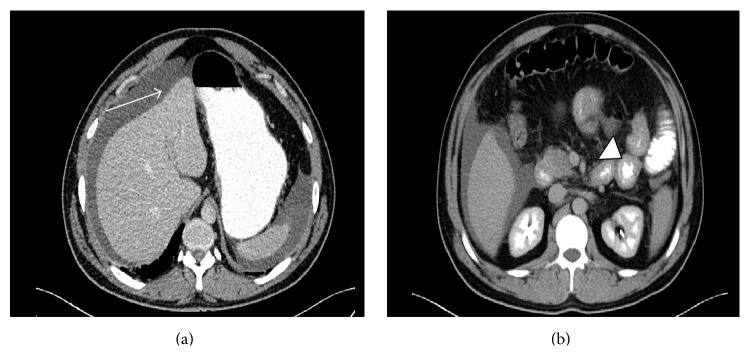
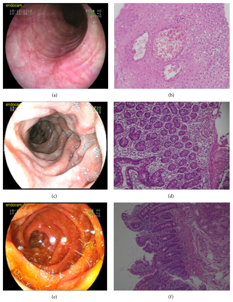
Eosinophilic gastroenteritis is a rare disorder of unknown cause characterized by focal or diffuse eosinophilic infiltration of gastrointestinal tract, especially the stomach and duodenum. Its clinical presentation depends on which segment of gastrointestinal tract is affected and on the depth of eosinophilic infiltration in the affected segment. We present a case of a 35-year-old male with abdominal distention for six months. Laboratory testing revealed elevated eosinophil count and serum immunoglobulin E (IgE) levels. In abdominal tomography, ascites was observed, and eosinophilic infiltration was detected in duodenum biopsy samples, collected during endoscopic examination of upper gastrointestinal system. Clinical and pathologic findings of the patient responded to steroid dramatically. Even though their comorbidity is rare, eosinophilic gastroenteritis should be considered in differential diagnosis of patients with unspecified ascites.
Figures
Similar articles
-
Eosinophilic gastroenteritis with abdominal pain and ascites: A case report.World J Clin Cases. 2021 Jun 16;9(17):4238-4243. doi: 10.12998/wjcc.v9.i17.4238. World J Clin Cases. 2021. PMID: 34141786 Free PMC article.
-
Eosinophilic ascites: A case report and literature review.J Family Community Med. 2015 Sep-Dec;22(3):183-5. doi: 10.4103/2230-8229.163042. J Family Community Med. 2015. PMID: 26392801 Free PMC article.
-
Eosinophilic ascites as an uncommon presentation of eosinophilic gastroenteritis: A case report.Arab J Gastroenterol. 2021 Jun;22(2):184-186. doi: 10.1016/j.ajg.2021.02.002. Epub 2021 Jun 2. Arab J Gastroenterol. 2021. PMID: 34090834
-
Eosinophilic gastroenteritis manifesting with ascites.South Med J. 1994 Sep;87(9):956-7. doi: 10.1097/00007611-199409000-00021. South Med J. 1994. PMID: 8091267 Review.
-
Eosinophilic gastroenteritis presenting as small bowel obstruction: a case report and review of the literature.World J Gastroenterol. 2007 Mar 21;13(11):1758-60. doi: 10.3748/wjg.v13.i11.1758. World J Gastroenterol. 2007. PMID: 17461485 Free PMC article. Review.
Cited by
-
A rare case of eosinophilic esophagitis and eosinophilic subserosal gastroenteritis with ascites.Turk J Gastroenterol. 2019 Sep;30(9):851-853. doi: 10.5152/tjg.2019.18680. Turk J Gastroenterol. 2019. PMID: 31530531 Free PMC article. No abstract available.
References
-
- Klein N. C., Hargrove R. L., Sleisenger M. H., Jeffries G. H. Eosinophilic gastroenteritis. Medicine. 1970;49(4):299–319. - PubMed
LinkOut - more resources
Full Text Sources
Other Literature Sources
