Use of an Ultrasonic Osteotome for Direct Removal of Beak-Type Ossification of Posterior Longitudinal Ligament in the Thoracic Spine
- PMID: 26819697
- PMCID: PMC4728100
- DOI: 10.3340/jkns.2015.58.6.571
Use of an Ultrasonic Osteotome for Direct Removal of Beak-Type Ossification of Posterior Longitudinal Ligament in the Thoracic Spine
Abstract
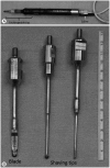
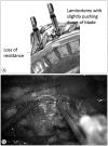
Direct removal of beak-type ossification of posterior longitudinal ligament at thoracic spine (T-OPLL) is a challenging surgical technique due to the potential risk of neural injury. Slipping off the cutting surface of a high-speed drill may result in entrapment in neural structures, leading to serious complications. Removal of T-OPLL with an ultrasonic osteotome, utilizing back and forth micro-motion of a blade rather than rotatory-motion of drill, may reduce such complications. We have applied the ultrasonic osteotome for posterior circumferential decompression of T-OPLL for three consecutive patients with beak-type OPLL and have described the surgical techniques and patient outcomes. The preoperative chief complaint was gait disturbance in all patients. Japanese orthopedic association scores (JOA) was used for functional assessment. Scores measured 2/11, 5/11, 2/11, and 4/11 for each patient. The ventral T-OPLL mass was exposed after posterior midline approach, laminotomy and transeversectomy. The T-OPLL mass was directly removed with an ultrasonic osteotome and instrumented segmental fixation was performed. The surgeries were uneventful. Detailed surgical techniques were presented. Gait disturbance was improved in all patients. Dural tear occurred in one patient without squeal. Postoperative JOA was 6/11, 10/11, 8/11, and 8/11 (recovery rate; 44%, 83%, 67%, and 43%) respectively at 18, 18, 10, and 1 months postoperative. T-OPLL was completely removed in all patients as confirmed with computed tomography scan. We hope that surgical difficulties in direct removal of T-OPLL might be reduced by utilizing ultrasonic osteotome.
Keywords: Beak; Ossification of posterior longitudinal ligament; Osteotome; Surgery; Thoracic spine; Ultrasound.
Figures


Similar articles
-
Gradual spinal cord decompression through migration of floated plaques after anterior decompression via a posterolateral approach for OPLL in the thoracic spine.J Neurosurg Spine. 2015 Oct;23(4):479-83. doi: 10.3171/2015.1.SPINE14960. Epub 2015 Jul 3. J Neurosurg Spine. 2015. PMID: 26140403
-
Minimally Invasive Anterior Decompression Technique without Instrumented Fusion for Huge Ossification of the Posterior Longitudinal Ligament in the Thoracic Spine: Technical Note And Literature Review.J Korean Neurosurg Soc. 2017 Sep;60(5):597-603. doi: 10.3340/jkns.2017.0404.017. Epub 2017 Aug 30. J Korean Neurosurg Soc. 2017. Retraction in: J Korean Neurosurg Soc. 2019 Sep;62(5):618. doi: 10.3340/jkns.2017.0404.017.r1. PMID: 28881124 Free PMC article. Retracted.
-
[Safety and effectiveness of ultrasonic osteotome in posterior cervical laminectomy decompression and fusion].Zhongguo Xiu Fu Chong Jian Wai Ke Za Zhi. 2018 Dec 15;32(12):1554-1559. doi: 10.7507/1002-1892.201804012. Zhongguo Xiu Fu Chong Jian Wai Ke Za Zhi. 2018. PMID: 30569683 Free PMC article. Chinese.
-
Surgical Treatment for Ossification of the Posterior Longitudinal Ligament (OPLL) at the Thoracic Spine: Usefulness of the Posterior Approach.Spine Surg Relat Res. 2018 Feb 28;2(3):169-176. doi: 10.22603/ssrr.2017-0044. eCollection 2018. Spine Surg Relat Res. 2018. PMID: 31440665 Free PMC article. Review.
-
Comparison of clinical outcomes in decompression and fusion versus decompression only in patients with ossification of the posterior longitudinal ligament: a meta-analysis.Neurosurg Focus. 2016 Jun;40(6):E9. doi: 10.3171/2016.3.FOCUS1630. Neurosurg Focus. 2016. PMID: 27246492
Cited by
-
Learning Curve and Clinical Outcomes of Ultrasonic Osteotome-based En Bloc Laminectomy for Thoracic Ossification of the Ligamentum Flavum.Orthop Surg. 2023 Sep;15(9):2318-2327. doi: 10.1111/os.13804. Epub 2023 Jul 5. Orthop Surg. 2023. PMID: 37403615 Free PMC article.
-
[Application of V-shaped stealth decompression technique using ultrasonic bone scalpel in anterior surgery for adjacent two-level cervical spondylosis].Zhongguo Xiu Fu Chong Jian Wai Ke Za Zhi. 2025 Jun 15;39(6):741-747. doi: 10.7507/1002-1892.202502056. Zhongguo Xiu Fu Chong Jian Wai Ke Za Zhi. 2025. PMID: 40545464 Free PMC article. Chinese.
-
Ultrasonic Bone Scalpel in Anterior Cervical Discectomy and Fusion Enhances Outcomes and Foraminal Decompression in Cervical Radiculopathy: A Retrospective Cohort Study.J Pain Res. 2025 Aug 4;18:3891-3902. doi: 10.2147/JPR.S525792. eCollection 2025. J Pain Res. 2025. PMID: 40799304 Free PMC article.
-
Ultrasonic bone scalpel for thoracic spinal decompression: case series and technical note.J Orthop Surg Res. 2020 Aug 8;15(1):309. doi: 10.1186/s13018-020-01838-9. J Orthop Surg Res. 2020. PMID: 32771031 Free PMC article.
-
Surgical Outcomes According to Dekyphosis in Patients with Ossification of the Posterior Longitudinal Ligament in the Thoracic Spine.J Korean Neurosurg Soc. 2020 Jan;63(1):89-98. doi: 10.3340/jkns.2018.0177. Epub 2019 May 14. J Korean Neurosurg Soc. 2020. PMID: 31079447 Free PMC article.
References
-
- Chaichana KL, Jallo GI, Dorafshar AH, Ahn ES. Novel use of an ultrasonic bone-cutting device for endoscopic-assisted craniosynostosis surgery. Childs Nerv Syst. 2013 [Epub ahead of print] - PubMed
-
- Fujimura Y, Nishi Y, Nakamura M, Toyama Y, Suzuki N. Long-term follow-up study of anterior decompression and fusion for thoracic myelopathy resulting from ossification of the posterior longitudinal ligament. Spine (Phila Pa 1976) 1997;22:305–311. - PubMed
-
- Hanai K, Ogikubo O, Miyashita T. Anterior decompression for myelopathy resulting from thoracic ossification of the posterior longitudinal ligament. Spine (Phila Pa 1976) 2002;27:1070–1076. - PubMed
Publication types
LinkOut - more resources
Full Text Sources
Other Literature Sources

