

Free Hand Insertion Technique of S2 Sacral Alar-Iliac Screws for Spino-Pelvic Fixation: Technical Note, Acadaveric Study
- PMID: 26819698
- PMCID: PMC4728101
- DOI: 10.3340/jkns.2015.58.6.578
Free Hand Insertion Technique of S2 Sacral Alar-Iliac Screws for Spino-Pelvic Fixation: Technical Note, Acadaveric Study
Abstract
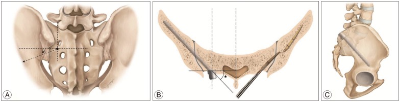
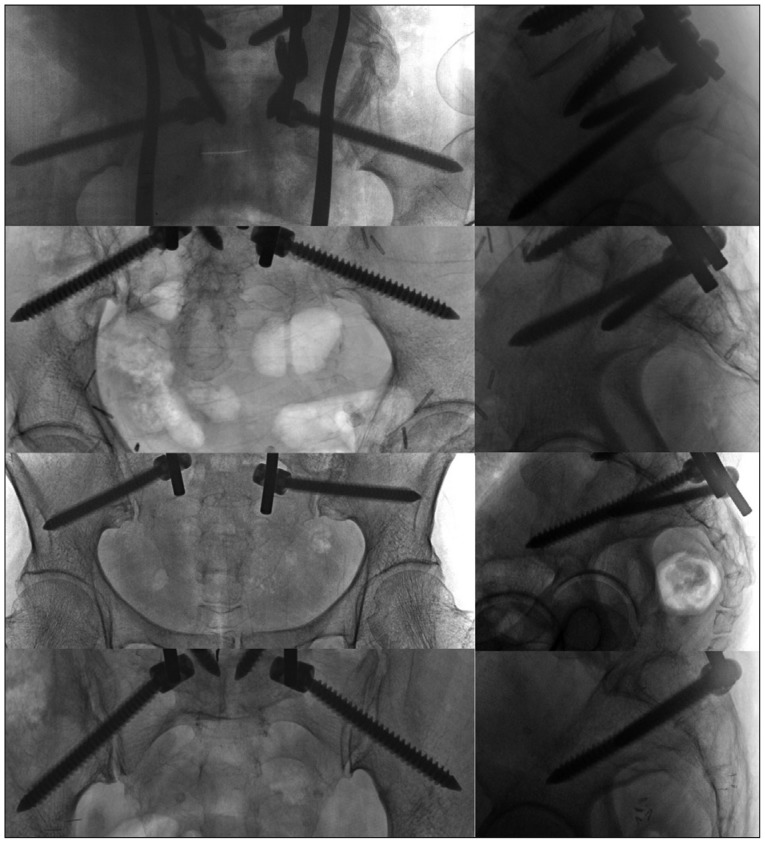
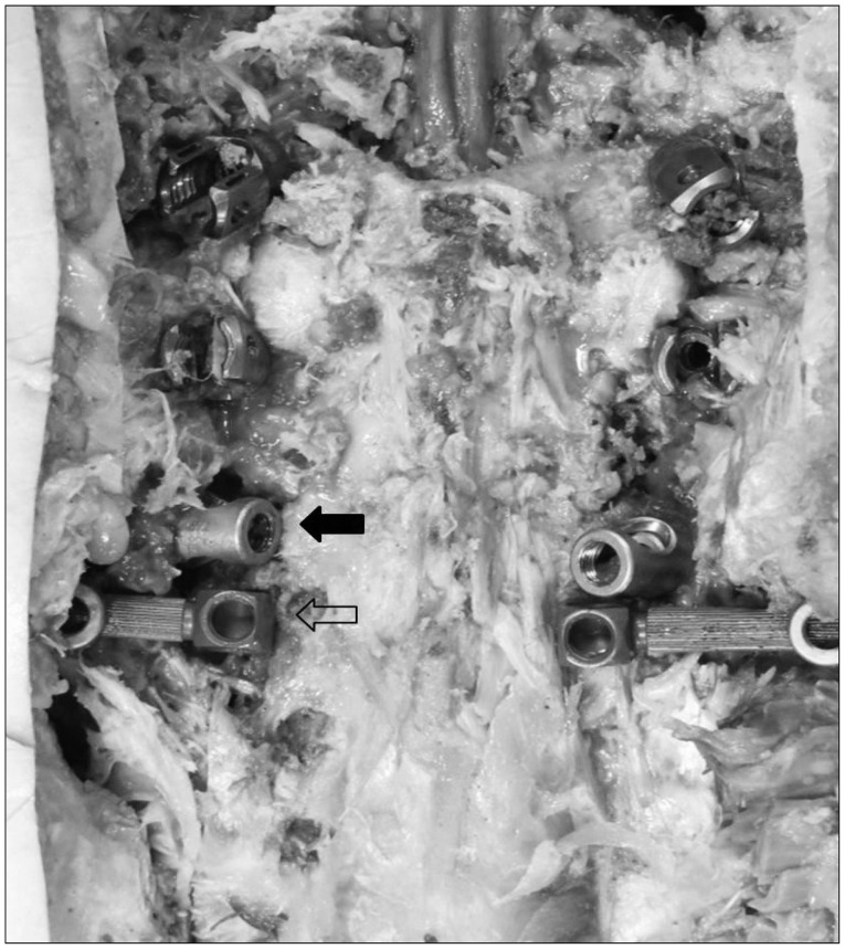
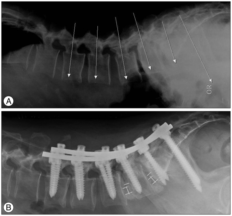
A rigid spino-pelvic fixation to anchor long constructs is crucial to maintain the stability of long fusion in spinal deformity surgery. Besides obtaining immediate stability and proper biomechanical strength of constructs, the S2 alar-iliac (S2AI) screws have some more advantages. Four Korean fresh-frozen human cadavers were procured. Free hand S2AI screw placement is performed using anatomic landmarks. The starting point of the S2AI screw is located at the midpoint between the S1 and S2 foramen and 2 mm medial to the lateral sacral crest. Gearshift was advanced from the desired starting point toward the sacro-iliac joint directing approximately 20° angulation caudally in sagittal plane and 30° angulation horizontally in the coronal plane connecting the posterior superior iliac spine (PSIS). We made a S2AI screw trajectory through the cancellous channel using the gearshift. We measured caudal angle in the sagittal plane and horizontal angle in the coronal plane. A total of eight S2AI screws were inserted in four cadavers. All screws inserted into the iliac crest were evaluated by C-arm and naked eye examination by two spine surgeons. Among 8 S2AI screws, all screws were accurately placed (100%). The average caudal angle in the sagittal plane was 17.3±5.4°. The average horizontal angle in the coronal plane connecting the PSIS was 32.0±1.8°. The placement of S2AI screws using the free hand technique without any radiographic guidance appears to an acceptable method of insertion without more radiation or time consuming.
Keywords: Adult deformity; Iliac fixation; S2 alar-iliac screw; Spino-pelvic fixation.
Figures
Similar articles
-
Multiple Points of Pelvic Fixation: Stacked S2-Alar-Iliac Screws (S2AI) or Concurrent S2AI and Open Sacroiliac Joint Fusion with Triangular Titanium Rod.JBJS Essent Surg Tech. 2022 Oct 7;12(4):e21.00044. doi: 10.2106/JBJS.ST.21.00044. eCollection 2022 Oct-Dec. JBJS Essent Surg Tech. 2022. PMID: 36743282 Free PMC article.
-
Low profile pelvic fixation: anatomic parameters for sacral alar-iliac fixation versus traditional iliac fixation.Spine (Phila Pa 1976). 2009 Mar 1;34(5):436-40. doi: 10.1097/BRS.0b013e318194128c. Spine (Phila Pa 1976). 2009. PMID: 19247163
-
The posterior superior iliac spine and sacral laminar slope: key anatomical landmarks for freehand S2-alar-iliac screw placement.J Neurosurg Spine. 2018 Oct;29(4):429-434. doi: 10.3171/2018.3.SPINE171374. Epub 2018 Jul 27. J Neurosurg Spine. 2018. PMID: 30052147
-
Robotic versus freehand S2 alar iliac fixation: in-depth technical considerations.J Spine Surg. 2018 Sep;4(3):638-644. doi: 10.21037/jss.2018.06.13. J Spine Surg. 2018. PMID: 30547130 Free PMC article. Review.
-
Postoperative complications of S2AI versus iliac screw in spinopelvic fixation: a meta-analysis and recent trends review.Spine J. 2020 Jun;20(6):964-972. doi: 10.1016/j.spinee.2019.11.014. Epub 2019 Dec 9. Spine J. 2020. PMID: 31830594 Review.
Cited by
-
Free-Hand Cervical Pedicle Screw Placement by Using Para-articular Minilaminotomy: Its Feasibility and Novice Neurosurgeons' Experience.Global Spine J. 2021 Jun;11(5):662-668. doi: 10.1177/2192568220919089. Epub 2020 Apr 30. Global Spine J. 2021. PMID: 32875896 Free PMC article.
-
Pelvic parameters directly influence ideal S2 alar-iliac (S2AI) screw trajectory.N Am Spine Soc J. 2020 Jul 12;2:100014. doi: 10.1016/j.xnsj.2020.100014. eCollection 2020 Aug. N Am Spine Soc J. 2020. PMID: 35141584 Free PMC article.
-
Freehand S2 Alar-Iliac Screw Placement Using K-Wire and Cannulated Screw : Technical Case Series.J Korean Neurosurg Soc. 2018 Jan;61(1):75-80. doi: 10.3340/jkns.2016.1212.008. Epub 2017 Dec 29. J Korean Neurosurg Soc. 2018. PMID: 29354238 Free PMC article.
-
A narrative review and scoring proposal for secondary lumbar instability after lumbar decompression surgery.Acta Neurochir (Wien). 2025 Jun 18;167(1):171. doi: 10.1007/s00701-025-06590-9. Acta Neurochir (Wien). 2025. PMID: 40531241 Free PMC article. Review.
-
Freehand S2-Alar-Iliac Screw Placement Technique in Lumbosacral Spinal Tumors: A Preliminary Study.Orthop Surg. 2022 Sep;14(9):2195-2202. doi: 10.1111/os.13434. Epub 2022 Aug 16. Orthop Surg. 2022. PMID: 35975359 Free PMC article.
References
-
- Chang TL, Sponseller PD, Kebaish KM, Fishman EK. Low profile pelvic fixation : anatomic parameters for sacral alar-iliac fixation versus traditional iliac fixation. Spine (Phila Pa 1976) 2009;34:436–440. - PubMed
-
- Edwards CC, 2nd, Bridwell KH, Patel A, Rinella AS, Berra A, Lenke LG. Long adult deformity fusions to L5 and the sacrum. A matched cohort analysis. Spine (Phila Pa 1976) 2004;29:1996–2005. - PubMed
-
- Kim YJ, Bridwell KH, Lenke LG, Cho KJ, Edwards CC, 2nd, Rinella AS. Pseudarthrosis in adult spinal deformity following multisegmental instrumentation and arthrodesis. J Bone Joint Surg Am. 2006;88:721–728. - PubMed
-
- Mattei TA, Fassett DR. Combined S-1 and S-2 sacral alar-iliac screws as a salvage technique for pelvic fixation after pseudarthrosis and lumbosacropelvic instability : combined S-1 and S-2 sacral alar-iliac screws as a salvage technique for pelvic fixation after pseudarthrosis and lumbosacropelvic instability : technical note. J Neurosurg Spine. 2013;19:321–330. - PubMed
LinkOut - more resources
Full Text Sources
Other Literature Sources
