

Reorganization of Synaptic Connections and Perineuronal Nets in the Deep Cerebellar Nuclei of Purkinje Cell Degeneration Mutant Mice
- PMID: 26819763
- PMCID: PMC4706924
- DOI: 10.1155/2016/2828536
Reorganization of Synaptic Connections and Perineuronal Nets in the Deep Cerebellar Nuclei of Purkinje Cell Degeneration Mutant Mice
Abstract
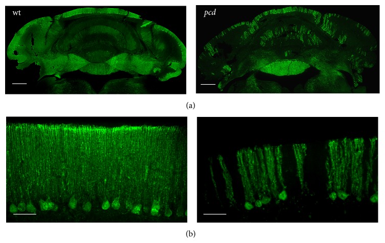
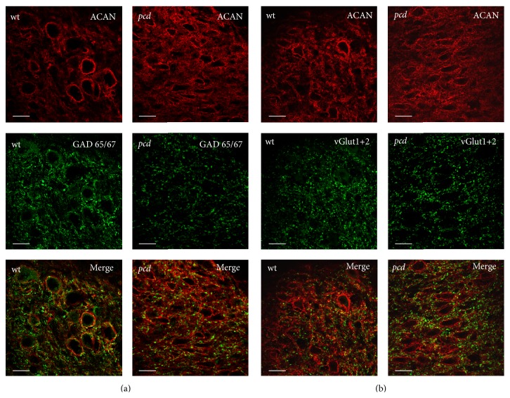
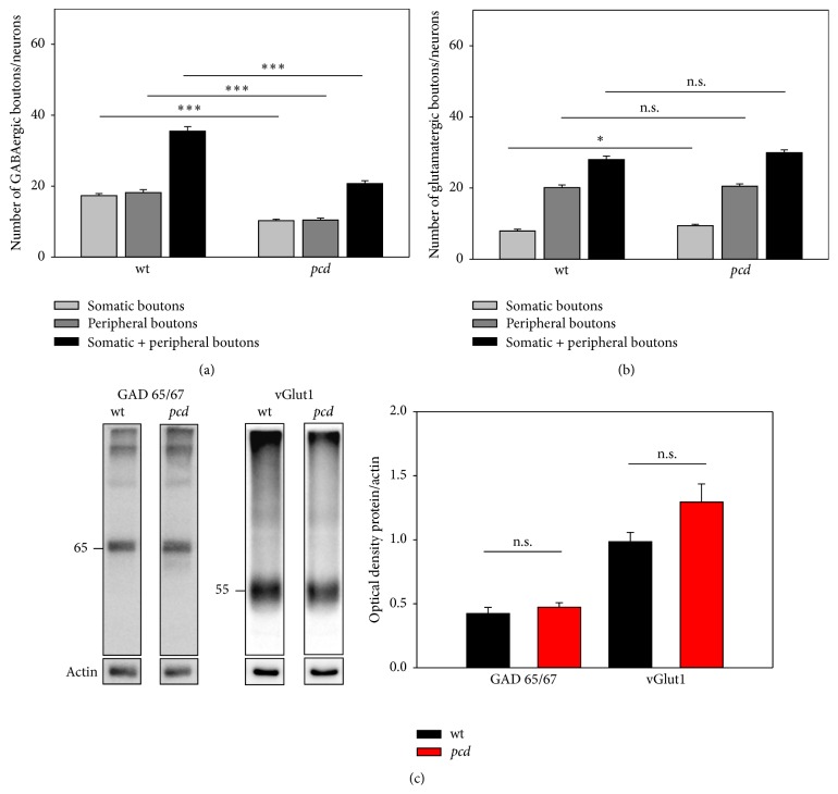
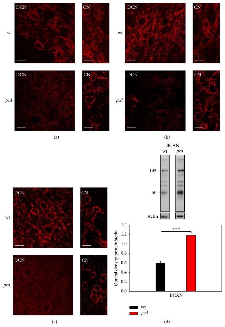
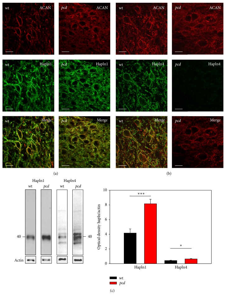
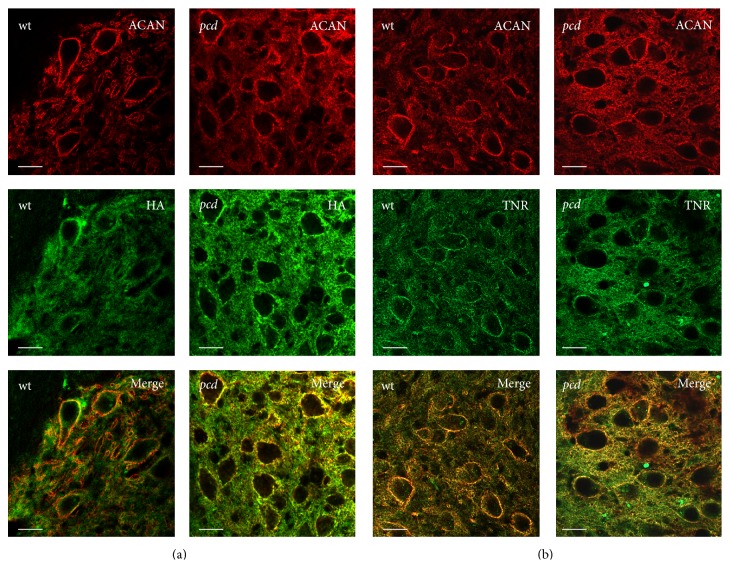
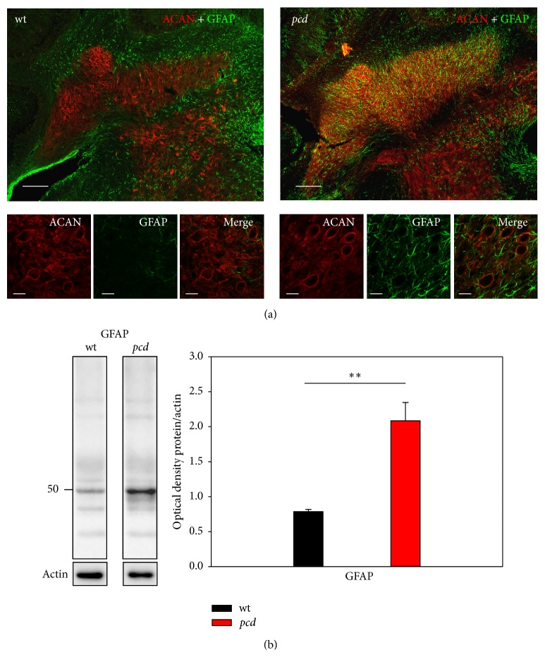
The perineuronal net (PN) is a subtype of extracellular matrix appearing as a net-like structure around distinct neurons throughout the whole CNS. PNs surround the soma, proximal dendrites, and the axonal initial segment embedding synaptic terminals on the neuronal surface. Different functions of the PNs are suggested which include support of synaptic stabilization, inhibition of axonal sprouting, and control of neuronal plasticity. A number of studies provide evidence that removing PNs or PN-components results in renewed neurite growth and synaptogenesis. In a mouse model for Purkinje cell degeneration, we examined the effect of deafferentation on synaptic remodeling and modulation of PNs in the deep cerebellar nuclei. We found reduced GABAergic, enhanced glutamatergic innervations at PN-associated neurons, and altered expression of the PN-components brevican and hapln4. These data refer to a direct interaction between ECM and synapses. The altered brevican expression induced by activated astrocytes could be required for an adequate regeneration by promoting neurite growth and synaptogenesis.
Figures
Similar articles
-
Perineuronal Nets in the Deep Cerebellar Nuclei Regulate GABAergic Transmission and Delay Eyeblink Conditioning.J Neurosci. 2018 Jul 4;38(27):6130-6144. doi: 10.1523/JNEUROSCI.3238-17.2018. Epub 2018 Jun 1. J Neurosci. 2018. PMID: 29858484 Free PMC article.
-
Experience-dependent plasticity and modulation of growth regulatory molecules at central synapses.PLoS One. 2011 Jan 31;6(1):e16666. doi: 10.1371/journal.pone.0016666. PLoS One. 2011. PMID: 21304956 Free PMC article.
-
Hapln4/Bral2 is a selective regulator for formation and transmission of GABAergic synapses between Purkinje and deep cerebellar nuclei neurons.J Neurochem. 2018 Dec;147(6):748-763. doi: 10.1111/jnc.14571. Epub 2018 Nov 11. J Neurochem. 2018. PMID: 30125937
-
Perineuronal Nets and Their Role in Synaptic Homeostasis.Int J Mol Sci. 2019 Aug 22;20(17):4108. doi: 10.3390/ijms20174108. Int J Mol Sci. 2019. PMID: 31443560 Free PMC article. Review.
-
Modulatory Effects of Monoamines and Perineuronal Nets on Output of Cerebellar Purkinje Cells.Front Neural Circuits. 2021 Jun 14;15:661899. doi: 10.3389/fncir.2021.661899. eCollection 2021. Front Neural Circuits. 2021. PMID: 34194302 Free PMC article. Review.
Cited by
-
Morphological and Functional Principles Governing the Plasticity Reserve in the Cerebellum: The Cortico-Deep Cerebellar Nuclei Loop Model.Biology (Basel). 2023 Nov 16;12(11):1435. doi: 10.3390/biology12111435. Biology (Basel). 2023. PMID: 37998034 Free PMC article.
-
The protein tyrosine phosphatase RPTPζ/phosphacan is critical for perineuronal net structure.J Biol Chem. 2020 Jan 24;295(4):955-968. doi: 10.1074/jbc.RA119.010830. Epub 2019 Dec 10. J Biol Chem. 2020. PMID: 31822561 Free PMC article.
-
A Gene-Expression Based Comparison of Murine and Human Inhibitory Interneurons in the Cerebellar Cortex and Nuclei.Cerebellum. 2025 Feb 28;24(2):55. doi: 10.1007/s12311-025-01809-y. Cerebellum. 2025. PMID: 40019676 Free PMC article.
-
Interactions between astrocytes and extracellular matrix structures contribute to neuroinflammation-associated epilepsy pathology.Front Mol Med. 2023 Jun 14;3:1198021. doi: 10.3389/fmmed.2023.1198021. eCollection 2023. Front Mol Med. 2023. PMID: 39086689 Free PMC article. Review.
-
Region-Specific Alterations of Perineuronal Net Expression in Postmortem Autism Brain Tissue.Front Mol Neurosci. 2022 Apr 13;15:838918. doi: 10.3389/fnmol.2022.838918. eCollection 2022. Front Mol Neurosci. 2022. PMID: 35493330 Free PMC article.
References
Publication types
MeSH terms
LinkOut - more resources
Full Text Sources
Other Literature Sources
Molecular Biology Databases
