Lateralizing Cortical Excitability in Drug Naïve Patients with Generalized or Focal Epilepsy
- PMID: 26819939
- PMCID: PMC4724855
- DOI: 10.14581/jer.15013
Lateralizing Cortical Excitability in Drug Naïve Patients with Generalized or Focal Epilepsy
Abstract
Background and purpose: Numerous transcranial magnetic stimulation (TMS) studies have defined the characteristic features of TMS in epilepsy. TME parameters were expected to classify the epilepsy syndrome or drug responses. However, the results such as cortical silent periods (CSP) are variable according to conditions of patients. Here, we investigate whether specific TMS parameters have localizing or lateralizing values in drug-naïve epilepsy patients.
Methods: We recruited 148 consecutive untreated patients with epilepsy (idiopathic generalized epilepsy (IGE) 38, focal epilepsy (FE) 110, mean age 31.4 years) and 38 age- and gender-matched normal subjects. We obtained resting motor threshold (RMT), motor-evoked potential (MEP), CSP, short interval intracortical inhibition (SICI, inter-stimuli interval 2-5 ms), and intracortical facilitation (ICF, inter-stimuli interval 10-20 ms). TMS were performed during a seizure-free state of more than 48 h.
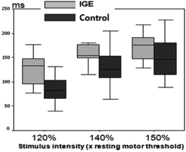
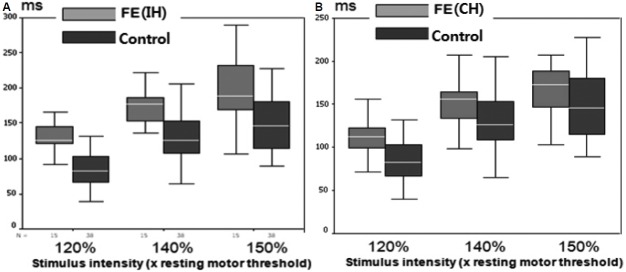
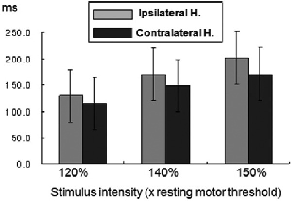
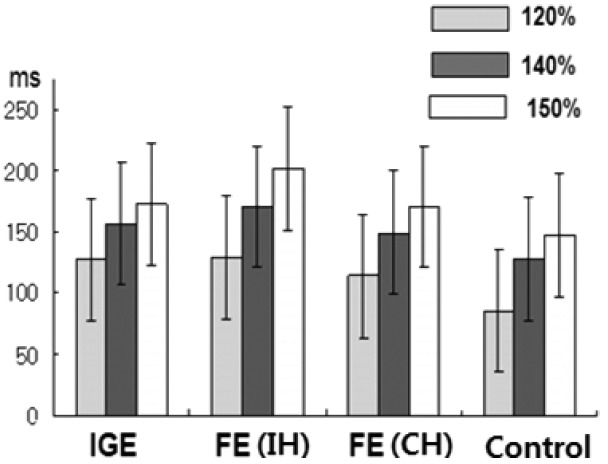
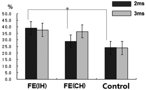
Results: In IGE, no interhemispheric difference in CSP was found (p > 0.05). However, the mean CSP was longer in IGE patients than in normal controls at all stimulus intensities (p < 0.05). The mean CSP in ipsilateral hemisphere (IH) of FE was significantly longer at all stimulus intensities than that in normal controls (p < 0.001). The CSP in IH was longer than that in the contralateral hemisphere of FE. There was no significant difference in CSP between FE and IGE. SICI was significantly reduced only in the IH of FE versus normal subjects. RMT, MEP amplitudes, and ICF did not differ among IGE, FE, and normal controls.
Conclusions: We found that prolonged CSP and reduced SICI in FE indicate asymmetrically increased cortical inhibition and excitation in the epileptic hemispheres. It suggests that CSP among TMS parameters has a crucial role to lateralize the epileptic hemisphere in FE.
Keywords: Cortical silent period; Epilepsy; Intracortical inhibition; Transcranial magnetic stimulation.
Figures
Similar articles
-
Cortical excitability in drug naive juvenile myoclonic epilepsy.Seizure. 2013 Oct;22(8):662-9. doi: 10.1016/j.seizure.2013.05.001. Epub 2013 Jun 7. Seizure. 2013. PMID: 23746624
-
Recruitment of motor cortex inhibition differentiates between generalized and focal epilepsy.Epilepsy Res. 2009 Apr;84(2-3):210-6. doi: 10.1016/j.eplepsyres.2009.02.013. Epub 2009 Mar 14. Epilepsy Res. 2009. PMID: 19286351
-
Zonisamide decreases cortical excitability in patients with idiopathic generalized epilepsy.Clin Neurophysiol. 2008 Jun;119(6):1385-92. doi: 10.1016/j.clinph.2008.02.008. Epub 2008 Apr 8. Clin Neurophysiol. 2008. PMID: 18396455 Clinical Trial.
-
Single and paired pulse transcranial magnetic stimulation in drug naïve epilepsy.Clin Neurophysiol. 2016 Sep;127(9):3140-3155. doi: 10.1016/j.clinph.2016.06.025. Epub 2016 Jul 5. Clin Neurophysiol. 2016. PMID: 27472551 Review.
-
A Meta-analysis of the Cortical Silent Period in Epilepsies.Brain Stimul. 2015 Jul-Aug;8(4):693-701. doi: 10.1016/j.brs.2015.04.008. Epub 2015 Apr 24. Brain Stimul. 2015. PMID: 25981158 Review.
Cited by
-
Clinical diagnostic utility of transcranial magnetic stimulation in neurological disorders. Updated report of an IFCN committee.Clin Neurophysiol. 2023 Jun;150:131-175. doi: 10.1016/j.clinph.2023.03.010. Epub 2023 Mar 29. Clin Neurophysiol. 2023. PMID: 37068329 Free PMC article. Review.
References
-
- Engel J., Jr Inhibitory mechanisms of epileptic seizure generation. Adv Neurol. 1995;67:157–71. - PubMed
-
- Cantello R, Gianelli M, Civardi C, Mutani R. Magnetic brain stimulation: the silent period after the motor evoked potential. Neurology. 1992;42:1951–9. - PubMed
-
- Macdonell RA, Curatolo JM, Berkovic SF. Transcranial magnetic stimulation and epilepsy. J Clin Neurophysiol. 2002;19:294–306. - PubMed
-
- Schrader LM, Stern JM, Koski L, Nuwer MR, Engel J., Jr Seizure incidence during single- and paired-pulse transcranial magnetic stimulation (TMS) in individuals with epilepsy. Clin Neurophysiol. 2004;115:2728–37. - PubMed
-
- Tassinari CA, Cincotta M, Zaccara G, Michelucci R. Transcranial magnetic stimulation and epilepsy. Clin Neurophysiol. 2003;114:777–98. - PubMed
LinkOut - more resources
Full Text Sources
Other Literature Sources