

Targeted Mybpc3 Knock-Out Mice with Cardiac Hypertrophy Exhibit Structural Mitral Valve Abnormalities
- PMID: 26819945
- PMCID: PMC4725593
- DOI: 10.3390/jcdd2020048
Targeted Mybpc3 Knock-Out Mice with Cardiac Hypertrophy Exhibit Structural Mitral Valve Abnormalities
Abstract
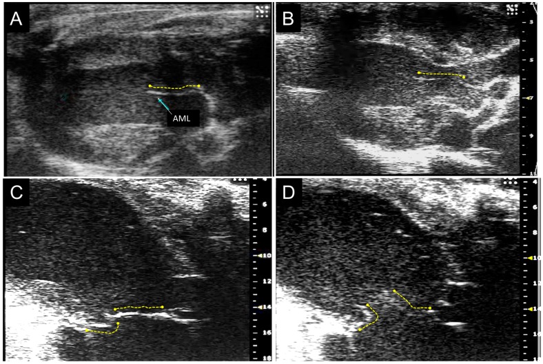
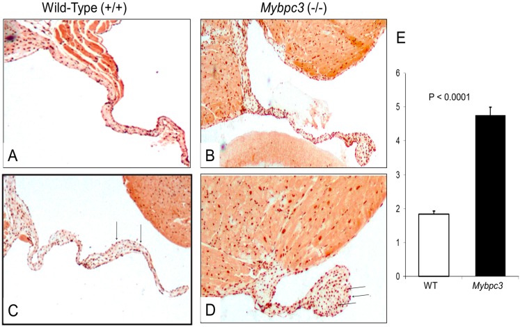
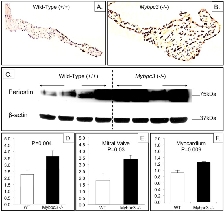
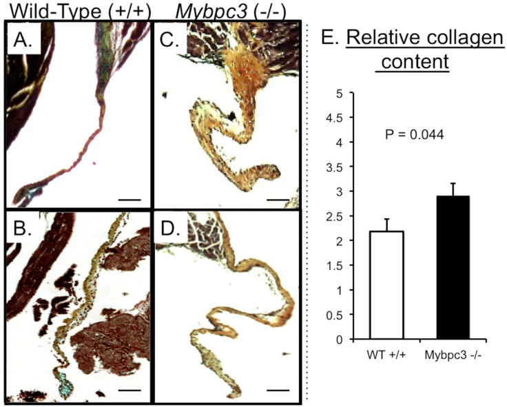
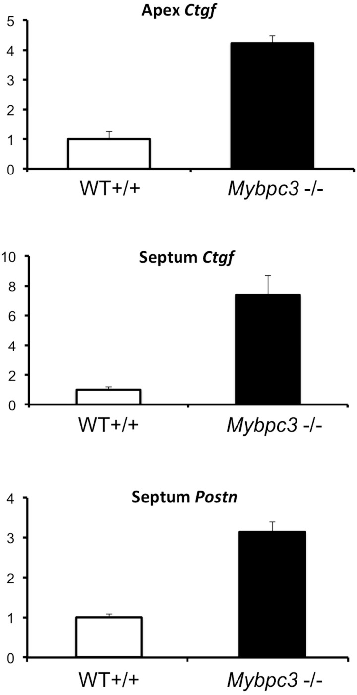
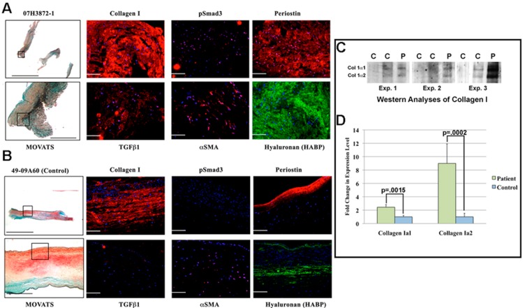
MYBPC3 mutations cause hypertrophic cardiomyopathy, which is frequently associated with mitral valve (MV) pathology. We reasoned that increased MV size is caused by localized growth factors with paracrine effects. We used high-resolution echocardiography to compare Mybpc3-null, heterozygous, and wild-type mice (n = 84, aged 3-6 months) and micro-CT for MV volume (n = 6, age 6 months). Mybpc3-null mice showed left ventricular hypertrophy, dilation, and systolic dysfunction compared to heterozygous and wild-type mice, but no systolic anterior motion of the MV or left ventricular outflow obstruction. Compared to wild-type mice, echocardiographic anterior leaflet length (adjusted for left ventricular size) was greatest in Mybpc3-null mice (1.92 ± 0.08 vs. 1.72 ± 0.08 mm, p < 0.001), as was combined leaflet thickness (0.23 ± 0.04 vs. 0.15 ± 0.02 mm, p < 0.001). Micro-CT analyses of Mybpc3-null mice demonstrated increased MV volume (0.47 ± 0.06 vs. 0.15 ± 0.06 mm3, p = 0.018) and thickness (0.35 ± 0.04 vs. 0.12 ± 0.04 mm, p = 0.002), coincident with increased markers of TGFβ activity compared to heterozygous and wild-type littermates. Similarly, excised MV from a patient with MYBPC3 mutation showed increased TGFβ activity. We conclude that MYBPC3 deficiency causes hypertrophic cardiomyopathy with increased MV leaflet length and thickness despite the absence of left ventricular outflow-tract obstruction, in parallel with increased TGFβ activity. MV changes in hypertrophic cardiomyopathy may be due to paracrine effects, which represent targets for therapeutic studies.
Keywords: TGFβ; hypertrophic cardiomyopathy; mitral valve.
Conflict of interest statement
The authors declare no conflict of interest.
Figures
References
-
- Hagege A.A., Dubourg O., Desnos M., Mirochnik R., Isnard G., Bonne G., Carrier L., Guicheney P., Bouhour J.B., Schwartz K., Komajda M. Familial hypertrophic cardiomyopathy. Cardiac ultrasonic abnormalities in genetically affected subjects without echocardiographic evidence of left ventricular hypertrophy. Eur. Heart J. 1998;19:490–499. doi: 10.1053/euhj.1997.0735. - DOI - PubMed
-
- Maron M.S., Olivotto I., Harrigan C., Appelbaum E., Gibson C.M., Lesser J.R., Haas T.S., Udelson J.E., Manning W.J., Maron B.J. Mitral Valve Abnormalities Identified by Cardiovascular Magnetic Resonance Represent a Primary Phenotypic Expression of Hypertrophic Cardiomyopathy. Circulation. 2011;124:40–47. doi: 10.1161/CIRCULATIONAHA.110.985812. - DOI - PubMed
Grants and funding
LinkOut - more resources
Full Text Sources
Other Literature Sources
Molecular Biology Databases