. 2016 Jan 21;3(1):e187.
doi: 10.1212/NXI.0000000000000187. eCollection 2016 Feb.

Mycoplasma pneumoniae, a trigger for Weston Hurst syndrome

Affiliations

Mycoplasma pneumoniae, a trigger for Weston Hurst syndrome

Rick Magun et al. Neurol Neuroimmunol Neuroinflamm. .

Abstract

Objective: We report a case of Mycoplasma pneumoniae infection as one possible trigger for Weston Hurst syndrome (acute hemorrhagic leukoencephalitis), a rare disorder of microvascular injury often described as a postinfectious complication of an upper respiratory illness.

Methods: This is a case of a 27-year-old man presenting with a Glasgow Coma Scale score of 3 and an acute head CT revealing extensive vasogenic edema in the right hemisphere associated with mass effect in the context of a recent upper respiratory illness. Right frontal biopsy was performed on day 2, which showed acute cerebritis, and the patient was aggressively treated with antibiotics. However, over the next 5 days from presentation, the vasogenic edema increased, leading ultimately to brain herniation and death.

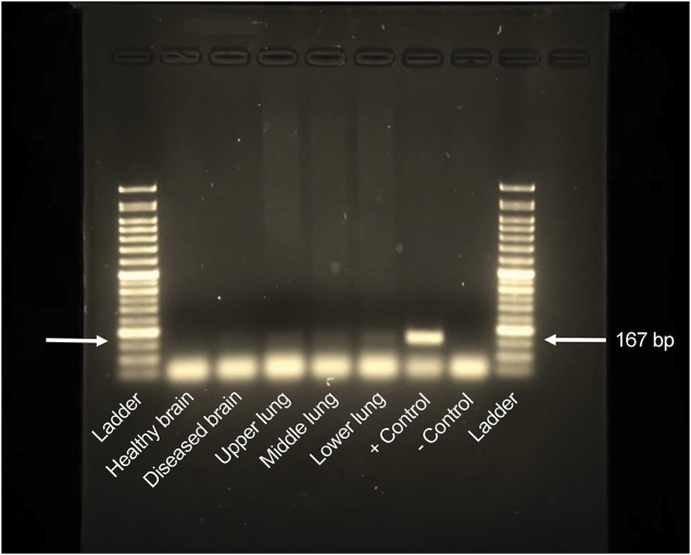
Results: A full autopsy was performed at 5 days from presentation, which showed areas of vessel wall fibrinoid necrosis throughout the right hemisphere as well as, but less so, in the left frontal lobe and pons. Chest x-ray on presentation revealed atypical pneumonia, blood tests were positive for cold agglutinins, and at full autopsy, there was myocarditis, all in keeping with recent M pneumoniae infection. DNA obtained from lung and diseased brain (postmortem) was positive for Mycoplasma providing more direct evidence for brain invasion by this organism as the ultimate trigger for Weston Hurst syndrome.

Conclusions: This is a rare case report of Weston Hurst syndrome having both initial brain biopsy on day 2 and full autopsy results on day 5 of presentation revealing important clinical clues about the pathogenesis of this often fatal disorder.


Figures

Figure 1
Figure 1. Summary of clinical presentation
(A) Head CT on days 1 and 2 showed extensive vasogenic edema in the right temporal-parietal region, a focal hypodensity in the right frontal region associated with mass effect, and subfalcine herniation to the left with early central downward herniation. Therefore, an urgent right-sided craniectomy was performed to relieve the pressure and obtain a biopsy of the right frontal region. (B) Chest x-ray on day 2 showed patchy congestion consistent with a right lower lobe pneumonia as well as pulmonary edema. Later, autopsy results at day 5 confirmed these findings and also showed mucoid purulent secretions in the bronchial tree. Myocarditis was also noted at autopsy with a primary neutrophilic reaction. Nasopharyngeal swab was negative for enteroviruses and respiratory viruses via PCR. Cold agglutinins were positive at a titer of 64 when tested against adult cells at 4°C.
Figure 2
Figure 2. Gross autopsy findings
A full Autopsy was performed after the patient died on Day 5 and the brain weight was 1600 grams. There is evidence of a previous right frontal biopsy site. On external examination of the brain there was hemorrhagic necrosis and swelling noted along the right lateral edge of the frontal/temporal/parietal area (arrows, A, C–F, H) with evidence of flattening of the right olfactory tract secondary to increase intracranial pressure (arrow, B). There was subfalcine herniation to the left, with herniation of the cingulate gyrus underneath the edge of the falx cerebri (arrow, G). There was also transtentorial herniation on the right with early signs of Duret's hemorrhages in the pons (arrows, I, J). A normal pons is shown for comparison (I). There was also cerebellar tonsillar herniation noted on the right which was likely the fatal complication.
Figure 3
Figure 3. Initial brain biopsy and autopsy microscopic findings
Initial brain biopsy on day 2 of the right frontal region showed acute cerebritis. There was a marked acute inflammatory cell reaction most consistent with an acute infectious process noted in lower (A) and higher (B) magnification. The neutrophil predominance was determined to be consistent with a pyogenic process, although the Gram stain was negative. Silver stain was also negative for fungus. There was no neoplasm, granulomatous inflammation, or vasculitis present. (C, D) Hematoxylin & eosin stain at autopsy showed punctate, ring-shaped foci of perivascular hemorrhage and edema, localized to the white matter. This was surrounded by inflammation with a predominantly macrophage reaction in the right and left orbital frontal areas as well as right frontal-parietal sections. (E, F) Trichrome stain confirmed fibrinoid necrosis in multiple vessels in the right orbital frontal and frontal-parietal areas. There was no lymphocytic infiltration and no microglial nodules as seen in vasculitis and viral encephalitis, respectively. There was no inflammation in the vessel walls. Thromboemboli, viral cytopathologic effects, foreign abscesses, and granulomas were not seen.
Figure 4
Figure 4. Autopsy immunochemistry results
CD68 (A) and ferritin (B) stain showed the presence of scattered microglial cells, foamy macrophages, and rare hemosiderin-laden macrophages. CD4 was negative for T cells (C). Other tests performed include staining T cell markers CD3, CD4, CD5, and CD8, as well as B cell marker CD20 (data not shown), all of which were negative. These results indicated a primarily macrophage reaction vs lymphocytic. Immunohistochemistry for immunoglobulin G (IgG) and IgM showed a perivascular double ring-like effect consistent with an autoimmune inflammatory reaction or leakage (arrows, D). Immunohistochemistry for myelin basic protein (MBP) showed areas of reduced staining in a perivascular distribution throughout the white matter (E). The U fibers were spared. There was perivascular edema, gliosis, and mild axonal damage seen on glial fibrillary acidic protein (GFAP) (F) and neurofilament stain (G). Of note, neurofilament stain showed minimal axonal damage in contrast to the MBP stain, which was most consistent with demyelination. Herpes simplex virus 1 and 2 staining was negative. Gram stain and silver stain were also performed, which were negative for infection.
Figure 5
Figure 5. Autopsy Mycoplasma DNA analysis
DNA was isolated from the right hemisphere of the brain in an area of hemorrhage and normal brain from the autopsy of our patient as well as the right lung. PCR test for Mycoplasma DNA is shown above. A corresponding 167–base pair (bp) band, as seen in the control, is noted in all the right lung fields (upper, middle, and lower) and diseased brain consistent with amplification of Mycoplasma DNA. No band for Mycoplasma DNA was seen in normal areas of the brain. The results indicate a recent Mycoplasma pneumoniae infection with brain invasion, which ultimately led to Weston Hurst syndrome.

References

    1. Dos Santos MP, Martin J, Woulfe J, et al. Autopsy-proven acute hemorrhagic leukoencephalitis in an elderly patient. Can J Neurol Sci 2014;41:99–102. - PubMed
    1. Gillies CG, Grunnet M, Hamilton CW. Tubular inclusions in macrophages in the brain of a patient with acute hemorrhagic leukoencephalitis (Weston Hurst syndrome). Ultrastruct Pathol 1994;18:19–22. - PubMed
    1. Stamm B, Moschopulos M, Hungerbuehler H, et al. Neuroinvasion by Mycoplasma pneumoniae in acute disseminated encephalomyelitis. Emerg Infect Dis 2008;14:641–643. - PMC - PubMed
    1. Donnet A, Dufour H, Gambarelli D, et al. Acute Weston Hurst necrotizing hemorrhagic leukoencephalitis. Rev Neurol 1996;152:748–751. - PubMed
    1. Tshibanda L, Nchimi A, Otte M, et al. Hurst acute haemorrhagic leukoencephalitis: MRI findings [in French]. JBR-BTR 2007;90:290–293. - PubMed