Chronic sciatic nerve compression induces fibrosis in dorsal root ganglia
- PMID: 26820076
- PMCID: PMC4768999
- DOI: 10.3892/mmr.2016.4810
Chronic sciatic nerve compression induces fibrosis in dorsal root ganglia
Abstract
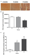
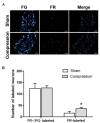
In the present study, pathological alterations in neurons of the dorsal root ganglia (DRG) were investigated in a rat model of chronic sciatic nerve compression. The rat model of chronic sciatic nerve compression was established by placing a 1 cm Silastic tube around the right sciatic nerve. Histological examination was performed via Masson's trichrome staining. DRG injury was assessed using Fluoro Ruby (FR) or Fluoro Gold (FG). The expression levels of target genes were examined using reverse transcription‑quantitative polymerase chain reaction, western blot and immunohistochemical analyses. At 3 weeks post‑compression, collagen fiber accumulation was observed in the ipsilateral area and, at 8 weeks, excessive collagen formation with muscle atrophy was observed. The collagen volume fraction gradually and significantly increased following sciatic nerve compression. In the model rats, the numbers of FR‑labeled DRG neurons were significantly higher, relative to the sham‑operated group, however, the numbers of FG‑labeled neurons were similar. In the ipsilateral DRG neurons of the model group, the levels of transforming growth factor‑β1 (TGF‑β1) and connective tissue growth factor (CTGF) were elevated and, surrounding the neurons, the levels of collagen type I were increased, compared with those in the contralateral DRG. In the ipsilateral DRG, chronic nerve compression was associated with significantly higher levels of phosphorylated (p)‑extracellular signal‑regulated kinase 1/2, and significantly lower levels of p‑c‑Jun N‑terminal kinase and p‑p38, compared with those in the contralateral DRGs. Chronic sciatic nerve compression likely induced DRG pathology by upregulating the expression levels of TGF‑β1, CTGF and collagen type I, with involvement of the mitogen‑activated protein kinase signaling pathway.
Figures


Similar articles
-
Chronic nerve compression injury induces a phenotypic switch of neurons within the dorsal root ganglia.J Comp Neurol. 2008 Jan 10;506(2):180-93. doi: 10.1002/cne.21537. J Comp Neurol. 2008. PMID: 18022951
-
[Expression of connective tissue growth factor in sciatic nerve after chronic compression injury and effect of rhodiola sachalinensis on its expression].Zhongguo Xiu Fu Chong Jian Wai Ke Za Zhi. 2014 Sep;28(9):1125-32. Zhongguo Xiu Fu Chong Jian Wai Ke Za Zhi. 2014. PMID: 25509779 Chinese.
-
Comparison of c-jun, junB, and junD mRNA expression and protein in the rat dorsal root ganglia following sciatic nerve transection.J Neurosci Res. 1995 Oct 15;42(3):391-401. doi: 10.1002/jnr.490420314. J Neurosci Res. 1995. PMID: 8583508
-
Changes in expression of PACAP in rat sensory neurons in response to sciatic nerve compression.Eur J Neurosci. 2004 Oct;20(7):1838-48. doi: 10.1111/j.1460-9568.2004.03644.x. Eur J Neurosci. 2004. PMID: 15380005
-
Chronic sciatic nerve compression secondary to arteriovenous malformation: case discussion and literature review.Ann R Coll Surg Engl. 2021 Oct;103(9):e278-e281. doi: 10.1308/rcsann.2020.7134. Epub 2021 Aug 25. Ann R Coll Surg Engl. 2021. PMID: 34431690 Free PMC article. Review.
Cited by
-
Chronic pain and local pain in usually painless conditions including neuroma may be due to compressive proximal neural lesion.Front Pain Res (Lausanne). 2023 Feb 20;4:1037376. doi: 10.3389/fpain.2023.1037376. eCollection 2023. Front Pain Res (Lausanne). 2023. PMID: 36890855 Free PMC article.
-
The Role of Endothelial Dysfunction in Peripheral Blood Nerve Barrier: Molecular Mechanisms and Pathophysiological Implications.Int J Mol Sci. 2019 Jun 20;20(12):3022. doi: 10.3390/ijms20123022. Int J Mol Sci. 2019. PMID: 31226852 Free PMC article. Review.
-
Tau protein function: The mechanical exploration of axonal transport disorder caused by persistent pressure in dorsal root ganglia.Mol Genet Genomic Med. 2019 Apr;7(4):e00580. doi: 10.1002/mgg3.580. Epub 2019 Jan 29. Mol Genet Genomic Med. 2019. PMID: 30697964 Free PMC article.
-
The ATP-P2X7 Signaling Pathway Participates in the Regulation of Slit1 Expression in Satellite Glial Cells.Front Cell Neurosci. 2019 Sep 19;13:420. doi: 10.3389/fncel.2019.00420. eCollection 2019. Front Cell Neurosci. 2019. PMID: 31607866 Free PMC article.
-
Coccydynia Improved by Percutaneous Discectomy.Cureus. 2024 Nov 27;16(11):e74564. doi: 10.7759/cureus.74564. eCollection 2024 Nov. Cureus. 2024. PMID: 39735113 Free PMC article.
References
Publication types
MeSH terms
Substances
LinkOut - more resources
Full Text Sources
Other Literature Sources
Research Materials
Miscellaneous

