. 2016 May;20(5):864-73.
doi: 10.1111/jcmm.12770. Epub 2016 Jan 28.

miR-655 suppresses epithelial-to-mesenchymal transition by targeting Prrx1 in triple-negative breast cancer

Affiliations

miR-655 suppresses epithelial-to-mesenchymal transition by targeting Prrx1 in triple-negative breast cancer

Zhi-Dong Lv et al. J Cell Mol Med. 2016 May.

Retraction in

Abstract

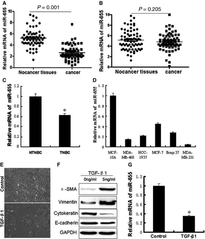
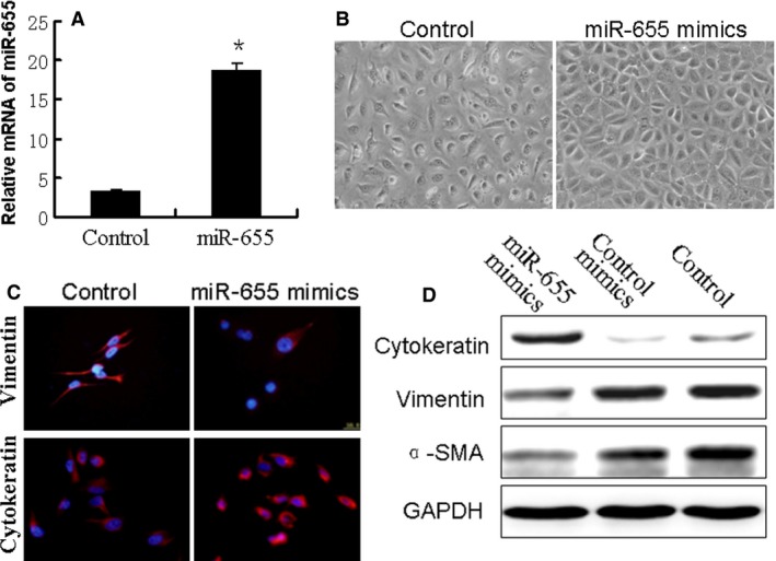
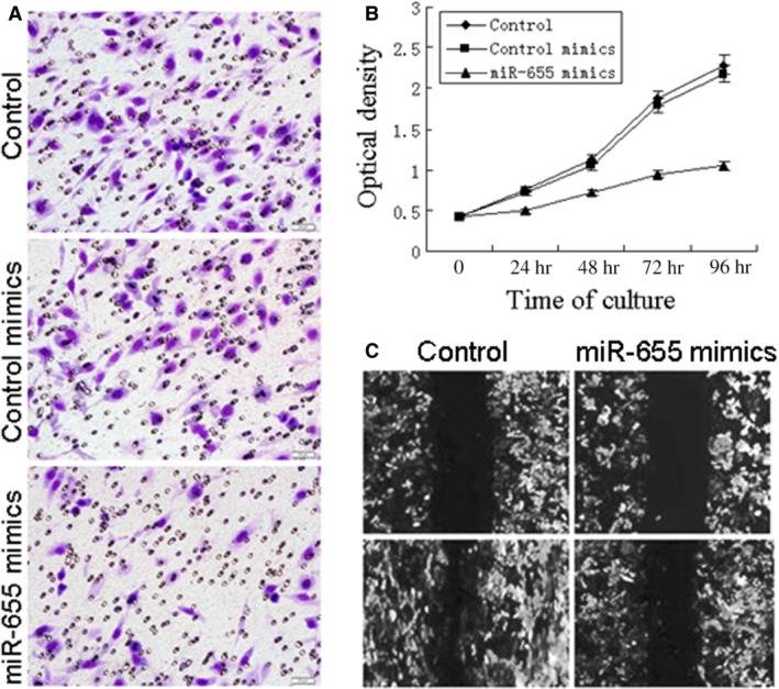
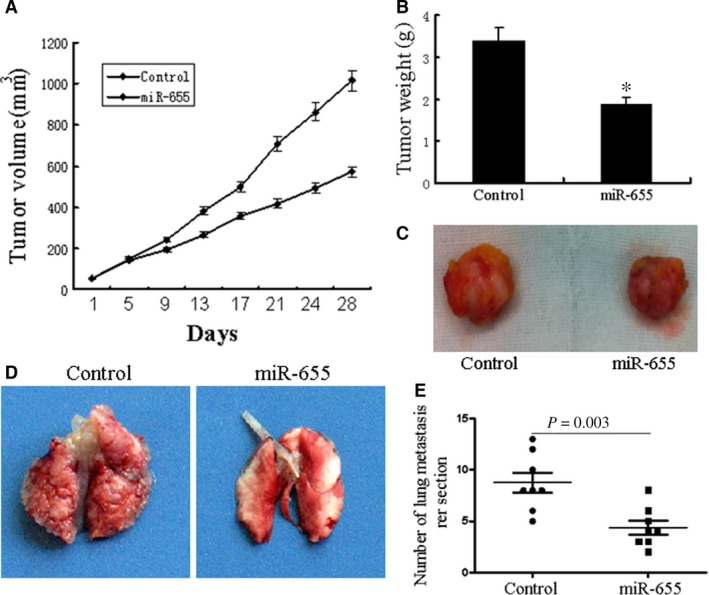
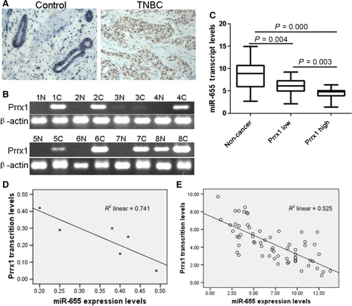
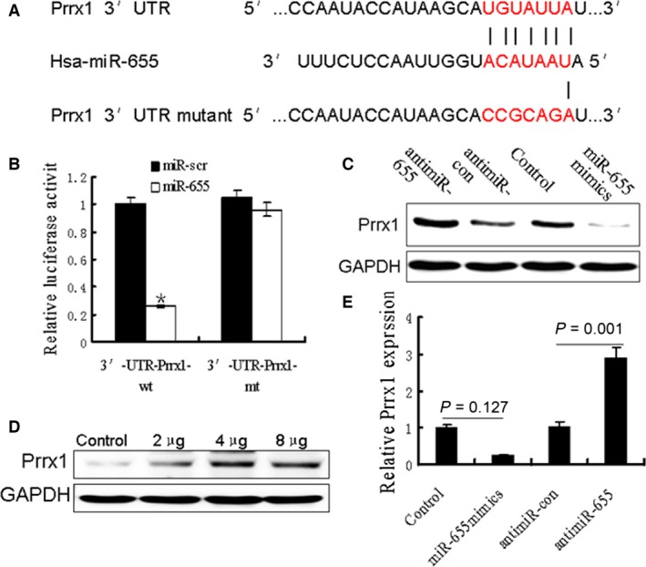
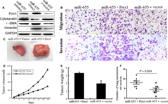
Triple-negative breast cancer (TNBC) is a highly aggressive breast cancer subtype that lacks effective targeted therapies. The epithelial-to-mesenchymal transition (EMT) is a key contributor in the metastatic process. In this study, we found that miR-655 was down-regulated in TNBC, and its expression levels were associated with molecular-based classification and lymph node metastasis in breast cancer. These findings led us to hypothesize that miR-655 overexpression may inhibit EMT and its associated traits of TNBC. Ectopic expression of miR-655 not only induced the up-regulation of cytokeratin and decreased vimentin expression but also suppressed migration and invasion of mesenchymal-like cancer cells accompanied by a morphological shift towards the epithelial phenotype. In addition, we found that miR-655 was negatively correlated with Prrx1 in cell lines and clinical samples. Overexpression of miR-655 significantly suppressed Prrx1, as demonstrated by Prrx1 3'-untranslated region luciferase report assay. Our study demonstrated that miR-655 inhibits the acquisition of the EMT phenotype in TNBC by down-regulating Prrx1, thereby inhibiting cell migration and invasion during cancer progression.

Keywords: Prrx1; epithelial-to-mesenchymal transition; miR-655; triple-negative breast cancer.


Figures

Figure 1
Figure 1
The miR‐655 expression levels were frequently down‐ regulated in TNBC and EMT model. (A) The levels of miR‐655 in 63 paired TNBC specimens and the corresponding paired normal adjacent tissues. (B) Expression levels of miR‐655 in 72 paired NTNBC specimens and the corresponding paired normal adjacent tissues. (C) Expression levels of miR‐655 in 63 TNBC specimens and 72 NTNBC specimens. (D) Expression levels of miR‐655 determined by qRTPCR in cells. Next, we establish a breast cancer EMT cell model by TGF‐β1, a well‐known EMT inducer. (E) Morphological differences in cancer cells treated with TGF‐β1 (40×). (F) Western blot analysis of the EMT markers' expression after TGF‐β1 treatment. (G) Screening of decreased miR‐655 by qRTPCR in the EMT model. All of the experiments were carried out in triplicate, and the results are displayed as the mean ± S.D., *P < 0.01.
Figure 2
Figure 2
Up‐regulation of miR‐655 altered the EMT phenotype in breast cancer cells. The breast cancer cells (MDAMB‐231) were transfected with miR‐655 mimics or negative control mimics for 48 hrs. (A) Overexpression of miR‐655 was validated with qRTPCR, *P < 0.05. (B) Morphological changes in miR‐655 overexpressed cells (100×). (C) Confocal immunofluorescence of cytokeratin and vimentin expression in MDAMB‐231 cells (100×). (D) Western blot analysis of cytokeratin, α‐SMA and vimentin expression in MDAMB‐231 cells.
Figure 3
Figure 3
Overexpression of miR‐655 suppress cell proliferation, migration, and invasion ability in vitro. The effects of miR‐655 on cell migration and invasion were detected using transwell chamber assays and wound healing assay. (A) Reduction in invasion caused by expression of miR‐655 (40×). (B) Ectopic expression of miR‐655 significantly suppressed the proliferation of breast cancer cells. (C) Overexpression of miR‐655 resulted in a significant decrease in migratory ability of MDAMB‐231 cells (40×).
Figure 4
Figure 4
miR‐655 inhibits TNBC growth and metastasis in vivo. Tumour growth in mouse xenograft models. MDAMB‐231 cells infected with miR‐655 lentivirus or scramble were injected subcutaneously into nude mice. (A) The tumour volume was measured every 4 days. After 28 days, the mice were killed. The photograph of excised tumours were performed (C), and the tumours were weighed (B). Tumour metastasis in mouse xenograft models. MDAMB‐231 cells overexpressing miR‐655 or scramble were injected into the tail vein of nude mice. After 60 days, the mice were killed. (D) The photograph of nude mice with lung dissemination from each group. (E) The disseminated nodules were evaluated. Each group had eight mice, *P < 0.05.
Figure 5
Figure 5
miR‐655 was negatively correlated with Prrx1 in cell lines and TNBC samples. (A) Immunohistochemistry results of Prrx1 expression in paired breast cancer tissue sample (40×)s. (B) RTPCR analysis demonstrated the Prrx1 expression in TNBC tissues and matched distal normal tissues. (C) miR‐655 was down‐regulated in breast cancer tissues. (D) Diagram represents the relative miR‐655 and Prrx1 mRNA expression levels in breast cancer cells. (E) In TNBC tissues, lower miR‐655 expression was accompanied by higher immunostaining of Prrx1.
Figure 6
Figure 6
Oncogene Prrx1 was specifically targeted by miR‐655. (A) The predicted binding sequences for miR‐655 within the human Prrx1 3′‐UTR. Seed sequences were highlighted and underlined. (B) Luciferase activity assays using a luciferase reporter with wild‐type or mutant human Prrx1 3′‐UTR were performed after co‐transfection of miR‐655 mimics or control into HEK293 cells. And mt 3′‐UTR had a significantly increase compared with wt 3′‐UTR. (C and E) Prrx1 expression was determined in breast cancer cells stably overexpressed miR‐655 and cells transfected with miR‐655 inhibitors, or anti‐miR‐control Western blot analysis. (D) Different concentrations of miR‐655 inhibitor transfection gradually increased Prrx1 expression in MDAMB‐231 cells.
Figure 7
Figure 7
Overexpression of Prrx1 reverse the inhibitory effects of miR‐655 on breast cancer cells. (A) Western blot analysed the Prrx1 and EMT‐related genes like α‐SMA, vimentin and cytokeratin in miR‐655‐vector cotransfected cells or miR‐655‐Prrx1 cotransfected cells compared with control group. (B) Transwell assay revealed that the reduction in migration and invasion caused by the overexpression of miR‐655 could be reversed by the introduction of Prrx1. Tumour growth in mouse xenograft models. miR‐655‐vector co‐transfected cells or miR‐655‐Prrx1 cotransfected MDAMB‐231 cells were injected subcutaneously into nude mice. (D) The tumour volume was measured every 4 days. After 28 days, the mice were killed. The photograph of excised tumours were performed (C), and the tumours were weighed (E). Tumour metastasis in mouse xenograft models. miR‐655‐vector co‐transfected cells or miR‐655‐Prrx1 cotransfected MDAMB‐231 cells were injected into the tail vein of nude mice. After 60 days, the mice were killed. (F) The disseminated nodules were evaluated. Each group had eight mice, *P < 0.01.

Similar articles

Cited by

References

    1. Baglia ML, Cai Q, Zheng Y, et al Dual specificity phosphatase 4 gene expression in association with triple‐negative breast cancer outcome. Breast Cancer Res Treat. 2014; 148: 211–20. - PMC - PubMed
    1. Lakshmaiah KC, Das U, Suresh TM, et al A study of triple negative breast cancer at a tertiary cancer care center in southern India. Ann Med Health Sci Res. 2014; 4: 933–7. - PMC - PubMed
    1. Guo W, Wang C, Guo Y, et al RASSF5A, a candidate tumor suppressor, is epigenetically inactivated in esophageal squamous cell carcinoma. Clin Exp Metastasis. 2015; 32: 83–98. - PubMed
    1. Xu XT, Tao ZZ, Song QB, et al SUZ12 RNA interference inhibits the invasion of gastric carcinoma cells. Hepatogastroenterology. 2014; 61: 2416–20. - PubMed
    1. Tian B, Li X, Kalita M, et al Analysis of the TGFβ‐induced program in primary airway epithelial cells shows essential role of NF‐κB/RelA signaling network in type II epithelial mesenchymal transition. BMC Genom. 2015; 16: 529. - PMC - PubMed

Publication types

MeSH terms