

Histone H1 Variants in Arabidopsis Are Subject to Numerous Post-Translational Modifications, Both Conserved and Previously Unknown in Histones, Suggesting Complex Functions of H1 in Plants
- PMID: 26820416
- PMCID: PMC4731575
- DOI: 10.1371/journal.pone.0147908
Histone H1 Variants in Arabidopsis Are Subject to Numerous Post-Translational Modifications, Both Conserved and Previously Unknown in Histones, Suggesting Complex Functions of H1 in Plants
Abstract
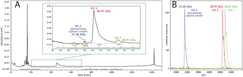
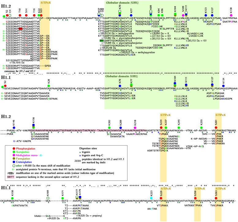
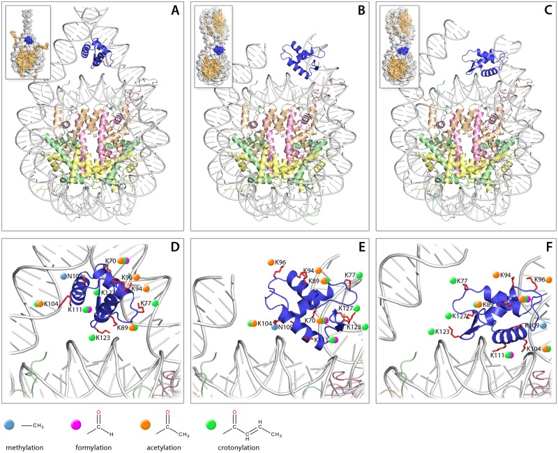
Linker histones (H1s) are conserved and ubiquitous structural components of eukaryotic chromatin. Multiple non-allelic variants of H1, which differ in their DNA/nucleosome binding properties, co-exist in animal and plant cells and have been implicated in the control of genetic programs during development and differentiation. Studies in mammals and Drosophila have revealed diverse post-translational modifications of H1s, most of which are of unknown function. So far, it is not known how this pattern compares with that of H1s from other major lineages of multicellular Eukaryotes. Here, we show that the two main H1variants of a model flowering plant Arabidopsis thaliana are subject to a rich and diverse array of post-translational modifications. The distribution of these modifications in the H1 molecule, especially in its globular domain (GH1), resembles that occurring in mammalian H1s, suggesting that their functional significance is likely to be conserved. While the majority of modifications detected in Arabidopsis H1s, including phosphorylation, acetylation, mono- and dimethylation, formylation, crotonylation and propionylation, have also been reported in H1s of other species, some others have not been previously identified in histones.
Conflict of interest statement
Figures

Similar articles
-
Distinctive core histone post-translational modification patterns in Arabidopsis thaliana.PLoS One. 2007 Nov 21;2(11):e1210. doi: 10.1371/journal.pone.0001210. PLoS One. 2007. PMID: 18030344 Free PMC article.
-
Combined bottom-up and top-down mass spectrometry analyses of the pattern of post-translational modifications of Drosophila melanogaster linker histone H1.J Proteomics. 2012 Jul 16;75(13):4124-38. doi: 10.1016/j.jprot.2012.05.034. Epub 2012 May 27. J Proteomics. 2012. PMID: 22647927
-
Histone H1 Purification and Post-Translational Modification Profiling by High-Resolution Mass Spectrometry.Methods Mol Biol. 2018;1675:147-166. doi: 10.1007/978-1-4939-7318-7_10. Methods Mol Biol. 2018. PMID: 29052191
-
Structure and function of histone methylation-binding proteins in plants.Biochem J. 2016 Jun 15;473(12):1663-80. doi: 10.1042/BCJ20160123. Biochem J. 2016. PMID: 27288029 Review.
-
Acetylation & Co: an expanding repertoire of histone acylations regulates chromatin and transcription.Essays Biochem. 2019 Apr 23;63(1):97-107. doi: 10.1042/EBC20180061. Print 2019 Apr 23. Essays Biochem. 2019. PMID: 30940741 Free PMC article. Review.
Cited by
-
Phylogeny-Based Systematization of Arabidopsis Proteins with Histone H1 Globular Domain.Plant Physiol. 2017 May;174(1):27-34. doi: 10.1104/pp.16.00214. Epub 2017 Mar 15. Plant Physiol. 2017. PMID: 28298478 Free PMC article.
-
Multiscale chromatin dynamics and high entropy in plant iPSC ancestors.J Cell Sci. 2024 Oct 15;137(20):jcs261703. doi: 10.1242/jcs.261703. Epub 2024 Jun 24. J Cell Sci. 2024. PMID: 38738286 Free PMC article.
-
Sperm-specific histone H1 in highly condensed sperm nucleus of Sargassum horneri.Sci Rep. 2024 Feb 9;14(1):3387. doi: 10.1038/s41598-024-53729-2. Sci Rep. 2024. PMID: 38336896 Free PMC article.
-
Salicylic acid interferes with GFP fluorescence in vivo.J Exp Bot. 2017 Mar 1;68(7):1689-1696. doi: 10.1093/jxb/erx031. J Exp Bot. 2017. PMID: 28369601 Free PMC article.
-
Histone Variants in the Specialization of Plant Chromatin.Annu Rev Plant Biol. 2022 May 20;73:149-172. doi: 10.1146/annurev-arplant-070221-050044. Epub 2022 Feb 15. Annu Rev Plant Biol. 2022. PMID: 35167758 Free PMC article. Review.
References
Publication types
MeSH terms
Substances
LinkOut - more resources
Full Text Sources
Other Literature Sources
