

Generation and characterization of human cardiac resident and non-resident mesenchymal stem cell
- PMID: 26820972
- PMCID: PMC5023579
- DOI: 10.1007/s10616-016-9946-5
Generation and characterization of human cardiac resident and non-resident mesenchymal stem cell
Abstract
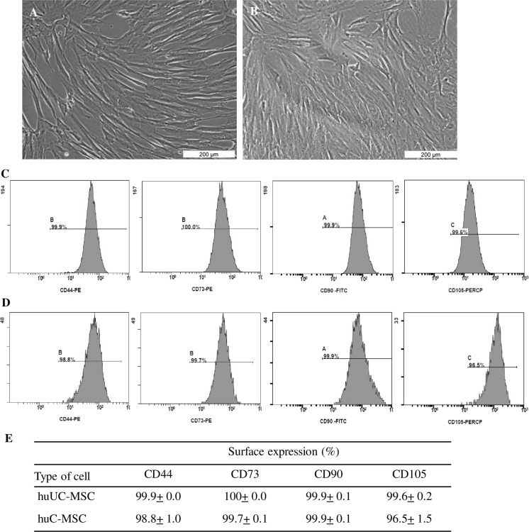
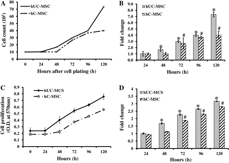
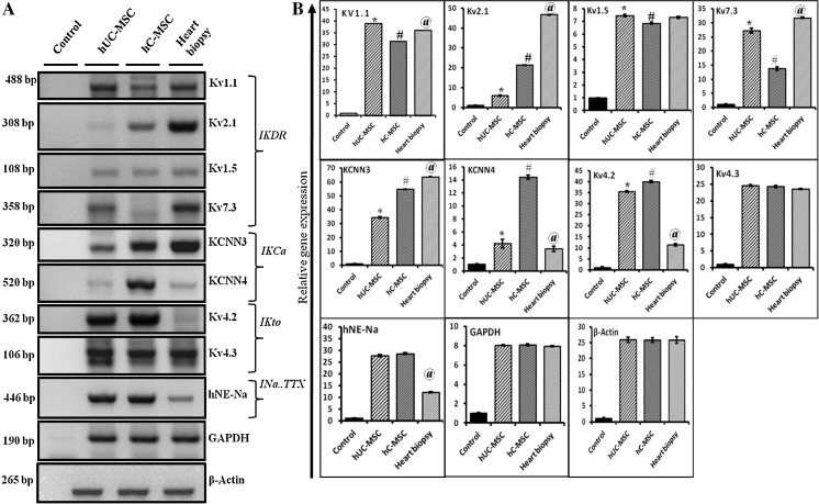
Despite the surgical and other insertional interventions, the complete recuperation of myocardial disorders is still elusive due to the insufficiency of functioning myocardiocytes. Thus, the use of stem cells to regenerate the affected region of heart becomes a prime important. In line with this human umbilical cord-derived mesenchymal stem cells (hUC-MSCs) have gained considerable interest due to their potential use for mesodermal cell based replacement therapy and tissue engineering. Since MSCs are harvested from various organs and anatomical locations of same organism, thus the cardiac regenerative potential of human cardiac-derived MSCs (hC-MSCs) and human umbilical cord Wharton's Jelly derived MSC (hUC-MSCs) were tested concurrently. At in vitro culture, both hUC-MSCs and hC-MSCs assumed spindle shape morphology with expression of typical MSC markers namely CD105, CD73, CD90 and CD44. Although, hUC-MSCs and hC-MSCs are identical in term of morphology and immunophenotype, yet hUC-MSCs harbored a higher cell growth as compared to the hC-MSCs. The inherent cardiac regenerative potential of both cells were further investigated with mRNA expression of ion channels. The RT-PCR results demonstrated that both MSCs were expressing a notable level of delayed rectifier-like K(+) current (I KDR ) ion channel, yet the relative expression level was considerably varied between hUC-MSCs and hC-MSCs that Kv1.1(39 ± 0.6 vs 31 ± 0.8), Kv2.1 (6 ± 0.2 vs 21 ± 0.12), Kv1.5 (7.4 ± 0.1 vs 6.8 ± 0.06) and Kv7.3 (27 ± 0.8 vs 13.8 ± 0.6). Similarly, the Ca2(+)-activated K(+) current (I KCa ) channel encoding gene, transient outward K(+) current (I to ) and TTX-sensitive transient inward sodium current (I Na.TTX ) encoding gene (Kv4.2, Kv4.3 and hNE-Na) expressions were detected in both groups as well. Despite the morphological and phenotypical similarity, the present study also confirms the existence of multiple functional ion channel currents IKDR, IKCa, Ito, and INa.TTX in undifferentiated hUC-MSCs as of hC-MSCs. Thus, the hUC-MSCs can be exploited as a potential candidate for future cardiac regeneration.
Keywords: Cardiac resident stem cell and umbilical cord stem cell; Electrophysiology; Mesenchymal stem cell.
Conflict of interest statement
The authors express no conflicts of interest towards the publication of this paper.
Figures
Similar articles
-
Impaired redox environment modulates cardiogenic and ion-channel gene expression in cardiac-resident and non-resident mesenchymal stem cells.Exp Biol Med (Maywood). 2017 Mar;242(6):645-656. doi: 10.1177/1535370216688568. Epub 2017 Jan 16. Exp Biol Med (Maywood). 2017. PMID: 28092181 Free PMC article.
-
Functional expression of ion channels in mesenchymal stem cells derived from umbilical cord vein.Stem Cells. 2007 Aug;25(8):2044-52. doi: 10.1634/stemcells.2006-0735. Epub 2007 May 24. Stem Cells. 2007. PMID: 17525238
-
Promising new potential for mesenchymal stem cells derived from human umbilical cord Wharton's jelly: sweat gland cell-like differentiative capacity.J Tissue Eng Regen Med. 2012 Aug;6(8):645-54. doi: 10.1002/term.468. Epub 2011 Sep 13. J Tissue Eng Regen Med. 2012. PMID: 21916019
-
Mitochondrial activity of human umbilical cord mesenchymal stem cells.Brain Circ. 2021 Mar 30;7(1):33-36. doi: 10.4103/bc.bc_15_21. eCollection 2021 Jan-Mar. Brain Circ. 2021. PMID: 34084975 Free PMC article. Review.
-
Mesenchymal stem cells in regenerative medicine: Focus on articular cartilage and intervertebral disc regeneration.Methods. 2016 Apr 15;99:69-80. doi: 10.1016/j.ymeth.2015.09.015. Epub 2015 Sep 15. Methods. 2016. PMID: 26384579 Review.
Cited by
-
Cardiac Nestin+ Mesenchymal Stromal Cells Enhance Healing of Ischemic Heart through Periostin-Mediated M2 Macrophage Polarization.Mol Ther. 2020 Mar 4;28(3):855-873. doi: 10.1016/j.ymthe.2020.01.011. Epub 2020 Jan 15. Mol Ther. 2020. PMID: 31991111 Free PMC article.
-
Impaired redox environment modulates cardiogenic and ion-channel gene expression in cardiac-resident and non-resident mesenchymal stem cells.Exp Biol Med (Maywood). 2017 Mar;242(6):645-656. doi: 10.1177/1535370216688568. Epub 2017 Jan 16. Exp Biol Med (Maywood). 2017. PMID: 28092181 Free PMC article.
-
Human Cardiac Mesenchymal Stem Cells Remodel in Disease and Can Regulate Arrhythmia Substrates.Circ Arrhythm Electrophysiol. 2020 Oct;13(10):e008740. doi: 10.1161/CIRCEP.120.008740. Epub 2020 Jul 29. Circ Arrhythm Electrophysiol. 2020. PMID: 32755466 Free PMC article.
-
Human Wharton's Jelly-Derived Mesenchymal Stem Cells Minimally Improve the Growth Kinetics and Cardiomyocyte Differentiation of Aged Murine Cardiac c-kit Cells in In Vitro without Rejuvenating Effect.Int J Mol Sci. 2019 Nov 6;20(22):5519. doi: 10.3390/ijms20225519. Int J Mol Sci. 2019. PMID: 31698679 Free PMC article.
-
Tissue engineering strategies for the induction of angiogenesis using biomaterials.J Biol Eng. 2018 Dec 27;12:36. doi: 10.1186/s13036-018-0133-4. eCollection 2018. J Biol Eng. 2018. PMID: 30603044 Free PMC article. Review.
References
-
- Bearzi C, Leri A, Lo Monaco F, Rota M, Gonzalez A, Hosoda T, Pepe M, Qanud K, Ojaimi C, Bardelli S, D’Amario D, D’Alessandro DA, Michler RE, Dimmeler S, Zeiher AM, Urbanek K, Hintze TH, Kajstura J, Anversa P. Identification of a coronary vascular progenitor cell in the human heart. Proc Natl Acad Sci USA. 2009;15:15885–15890. doi: 10.1073/pnas.0907622106. - DOI - PMC - PubMed
LinkOut - more resources
Full Text Sources
Other Literature Sources
Research Materials
Miscellaneous
