. 2016 Jan 28;7(1):e2073.
doi: 10.1038/cddis.2015.412.

X-linked inhibitor of apoptosis protein mediates tumor cell resistance to antibody-dependent cellular cytotoxicity

Affiliations

X-linked inhibitor of apoptosis protein mediates tumor cell resistance to antibody-dependent cellular cytotoxicity

M K Evans et al. Cell Death Dis. .

Abstract

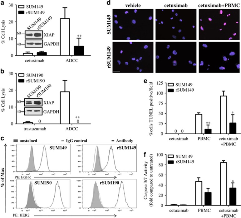
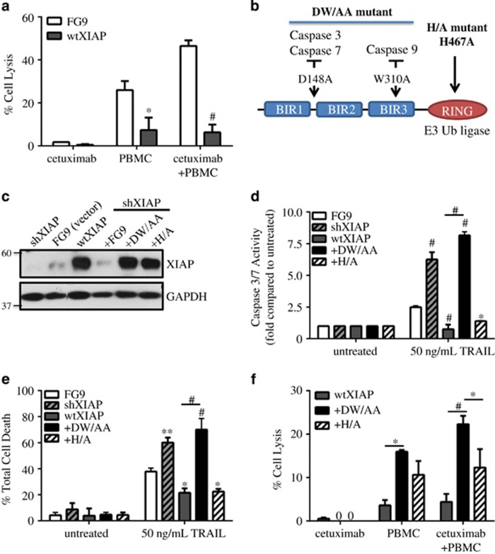
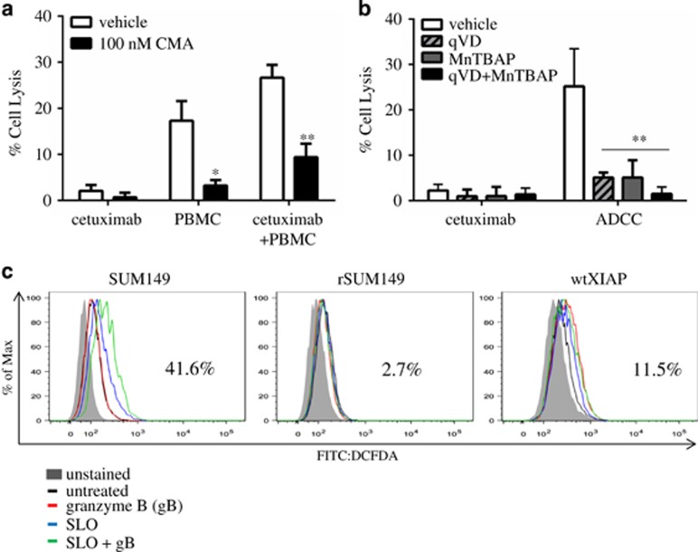
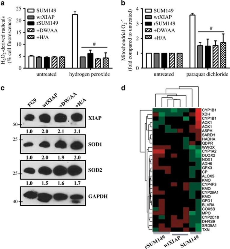
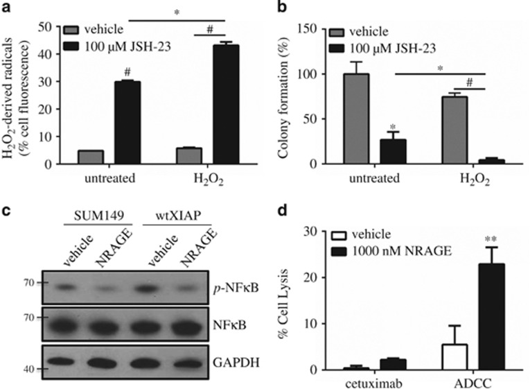
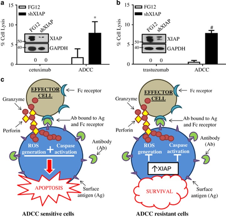
Inflammatory breast cancer (IBC) is the deadliest, distinct subtype of breast cancer. High expression of epidermal growth factor receptors [EGFR or human epidermal growth factor receptor 2 (HER2)] in IBC tumors has prompted trials of anti-EGFR/HER2 monoclonal antibodies to inhibit oncogenic signaling; however, de novo and acquired therapeutic resistance is common. Another critical function of these antibodies is to mediate antibody-dependent cellular cytotoxicity (ADCC), which enables immune effector cells to engage tumors and deliver granzymes, activating executioner caspases. We hypothesized that high expression of anti-apoptotic molecules in tumors would render them resistant to ADCC. Herein, we demonstrate that the most potent caspase inhibitor, X-linked inhibitor of apoptosis protein (XIAP), overexpressed in IBC, drives resistance to ADCC mediated by cetuximab (anti-EGFR) and trastuzumab (anti-HER2). Overexpression of XIAP in parental IBC cell lines enhances resistance to ADCC; conversely, targeted downregulation of XIAP in ADCC-resistant IBC cells renders them sensitive. As hypothesized, this ADCC resistance is in part a result of the ability of XIAP to inhibit caspase activity; however, we also unexpectedly found that resistance was dependent on XIAP-mediated, caspase-independent suppression of reactive oxygen species (ROS) accumulation, which otherwise occurs during ADCC. Transcriptome analysis supported these observations by revealing modulation of genes involved in immunosuppression and oxidative stress response in XIAP-overexpressing, ADCC-resistant cells. We conclude that XIAP is a critical modulator of ADCC responsiveness, operating through both caspase-dependent and -independent mechanisms. These results suggest that strategies targeting the effects of XIAP on caspase activation and ROS suppression have the potential to enhance the activity of monoclonal antibody-based immunotherapy.


Figures

Figure 1
Figure 1
Apoptotic dysregulation inhibits antibody-dependent cell cytotoxicity (ADCC) in breast cancer cells. (a) Percent cell lysis of SUM149 and rSUM149 cells incubated with cetuximab alone or ADCC conditions for 4 h, n=3–6. (b) Percent cell lysis of SUM190 and rSUM190 cells incubated with trastuzumab alone or ADCC conditions for 4 h. Bars represent mean±S.E.M. calculated percent lysis, n=3-4, **P<0.005. (c) Surface expression of EGFR in SUM149 and rSUM149 (top) and HER2 in SUM190 and rSUM190 (bottom) as measured by flow cytometry. Inset: Labeling of axes, representative of n=3 experiments. (d) TUNEL staining of SUM149 and rSUM149 cells treated with vehicle, cetuximab alone or cetuximab plus PBMCs. DAPI is shown in blue and TUNEL in red. Representative of n=2, magnification x40, scale bar=25 μm. (e) Quantification of the number of TUNEL-positive tumor cells from d. Bars represent %positive out of the total number of cells in each field/condition, n=3. (f) Caspase activity of SUM149 and rSUM149 cells cultured with cetuximab, PBMC alone or the combination for 4 h. Bars represent mean±S.E.M. fold relative light units compared with untreated, n=2–3. *P<0.05, **P<0.005 (comparison of rSUM149 to SUM149)
Figure 2
Figure 2
XIAP overexpression inhibits ADCC response in SUM149 cells through caspase inhibition. (a) Percent cell lysis of SUM149 FG9 and wtXIAP cells incubated with cetuximab alone, PBMC alone or cetuximab and IL-2 activated PBMCs for 4 h, n=2–3. (b) Schematic of XIAP mutants used in this study. D148A mutation is known to disrupt binding of caspases 3 and 7, whereas W310A disrupts caspase-9 binding. The H467A point mutation abolishes E3 ubiquitin ligase activity. (c) Western immunoblot of XIAP expression in cell lines transduced as indicated. FG9 is an empty vector as described in Materials and Methods. (d) Caspase activity and (e) viability of XIAP variant cell lines treated as indicated. Bars represent fold change in luminescence (d) or mean±S.E.M. % cell death (e), n=2–3. (f) Percent cell lysis of wtXIAP, +DW/AA and +H/A cells incubated with cetuximab alone, PBMC alone or cetuximab and IL-2 activated PBMCs for 4 h. Bars represent mean±S.E.M. calculated percent lysis, n=2–3. *P<0.05, **P<0.005, #P<0.001 (compared with FG9 unless otherwise indicated)
Figure 3
Figure 3
ADCC-mediated ROS accumulation is attenuated by exogenous antioxidants and XIAP overexpression. (a) Percent cell lysis of SUM149 cells incubated with cetuximab, PBMC alone or the combination in the presence or absence of concanamycin A (CMA), a perforin inhibitor. Bars represent mean±S.E.M. calculated percent lysis, n=2–3. (b) Percent cell lysis of SUM149 cells incubated with cetuximab alone or in ADCC conditions in the presence or absence of qVD (pan-caspase inhibitor), MnTBAP (antioxidant) or the combination. Bars represent mean±S.E.M. calculated percent lysis, n=2–3. *P<0.05 **P<0.005 (compared with vehicle). (c) Representative histograms of cells treated with: granzyme B (gB) alone (red line), streptolysin O (SLO) alone (blue line) or the combination of SLO+gB (green line) compared with untreated (black line). Number represents %positive for SLO+gB condition. Inset: Labeling of axes and diagram of sample colors, representative of n=2 experiments
Figure 4
Figure 4
XIAP overexpression inhibits ROS accumulation through upregulation of antioxidant capacity. (a) Fold induction of mitochondrial superoxides and (b) percentage of cells with high hydrogen peroxide-derived radicals in cells treated as indicated. Bars represent mean±S.E.M. relative to untreated cells. n=2–3, #P<0.001 (compared with SUM149). (c) Western immunoblot analysis of basal XIAP, SOD1 and SOD2 levels in SUM149 FG9, wtXIAP, +DW/AA and +H/A cells. Numbers represent densitometric analysis. (d) Normalized expression of the top most significantly differentially expressed genes shown as a heat map of over-(red) or under-(green) expressed genes in the oxidoreductase activity (GO:001649) GSEA category
Figure 5
Figure 5
NF-κB is essential for XIAP-mediated suppression of ROS and inhibition enhances ADCC. (a) Percentage of wtXIAP cells with high hydrogen peroxide-derived radicals in cells treated as indicated. Bars represent mean±S.E.M. relative to untreated cells, n=2–3. (b) Clonogenic growth assay in cells treated as indicated. Bars represent mean±S.E.M. colonies formed/cells plated as a percentage of the untreated sample, n=2–3. (c) Western immunoblotting for phospho-p65 (p-NF-κB), total p65 (NF-κB) and GAPDH as loading control in SUM149 and wtXIAP cells treated with vehicle or 1 μM NRAGE peptide. (d) Percent cell lysis of wtXIAP cells incubated with cetuximab alone or in ADCC conditions in the presence or absence of NRAGE peptide. Bars represent mean±S.E.M. calculated percent lysis, n=2–3. *P<0.05, **P<0.005, #P<0.001
Figure 6
Figure 6
Targeted inhibition of XIAP by RNAi sensitizes ADCC-resistant cells to apoptosis. Percent cell lysis of FG12 (control) or XIAP short hairpin RNA-transfected (a) rSUM149 cells and (b) rSUM190 cells incubated with antibody alone or ADCC conditions for 4 h. Bars represent mean±S.E.M. calculated percent lysis, n=4–5, *P<0.05, #P<0.001. Inset: Western immunoblot of XIAP expression at time of ADCC experiment. (c) Schematic of XIAP-mediated inhibition of ADCC. In ADCC-sensitive cells, antibody binding to surface antigen bridges tumor cells to effector cells, leading to subsequent release of lytic granules containing perforin and granzymes. Granzymes enter target cells through perforin channels, inducing both ROS generation and activating effector caspases leading to efficient apoptosis in tumor cells. In cells with XIAP overexpression, however, this process is abrogated through caspase-dependent and -independent mechanisms leading to tumor cell survival

References

    1. Robertson FM, Bondy M, Yang W, Yamauchi H, Wiggins S, Kamrudin S et al. Inflammatory breast cancer: the disease, the biology, the treatment. CA Cancer J Clin 2010; 60: 351–375. - PubMed
    1. Ueno NT, Buzdar AU, Singletary SE, Ames FC, McNeese MD, Holmes FA et al. Combined-modality treatment of inflammatory breast carcinoma: twenty years of experience at M. D. Anderson Cancer Center. Cancer Chemother Pharmacol 1997; 40: 321–329. - PubMed
    1. Prost S, Le MG, Douc-Rasy S, Ahomadegbe JC, Spielmann M, Guerin M et al. Association of c-erbB2-gene amplification with poor prognosis in non-inflammatory breast carcinomas but not in carcinomas of the inflammatory type. Int J Cancer 1994; 58: 763–768. - PubMed
    1. Van Laere SJ, Van der Auwera I, Van den Eynden GG, van Dam P, Van Marck EA, Vermeulen PB et al. NF-kappaB activation in inflammatory breast cancer is associated with oestrogen receptor downregulation, secondary to EGFR and/or ErbB2 overexpression and MAPK hyperactivation. Br J Cancer 2007; 97: 659–669. - PMC - PubMed
    1. Cabioglu N, Gong Y, Islam R, Broglio KR, Sneige N, Sahin A et al. Expression of growth factor and chemokine receptors: new insights in the biology of inflammatory breast cancer. Ann Oncol 2007; 18: 1021–1029. - PubMed

Publication types

MeSH terms