. 2016 Jan 29:7:10520.
doi: 10.1038/ncomms10520.

Reduction of aberrant NF-κB signalling ameliorates Rett syndrome phenotypes in Mecp2-null mice

Affiliations

Reduction of aberrant NF-κB signalling ameliorates Rett syndrome phenotypes in Mecp2-null mice

Noriyuki Kishi et al. Nat Commun. .

Abstract

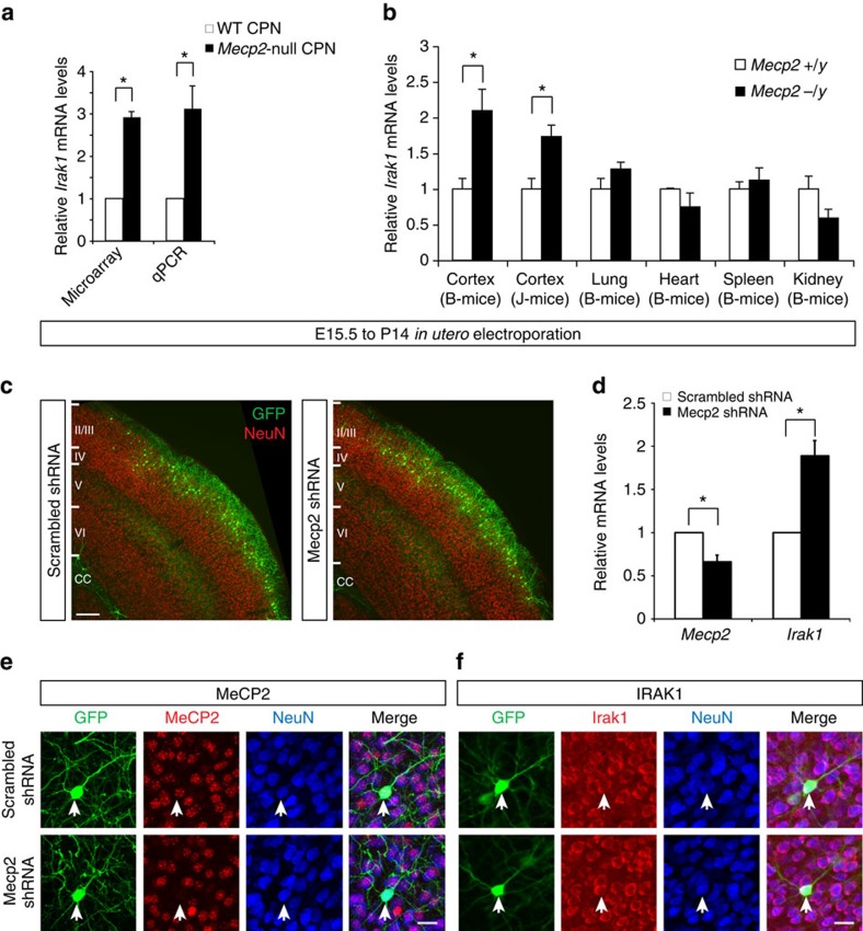
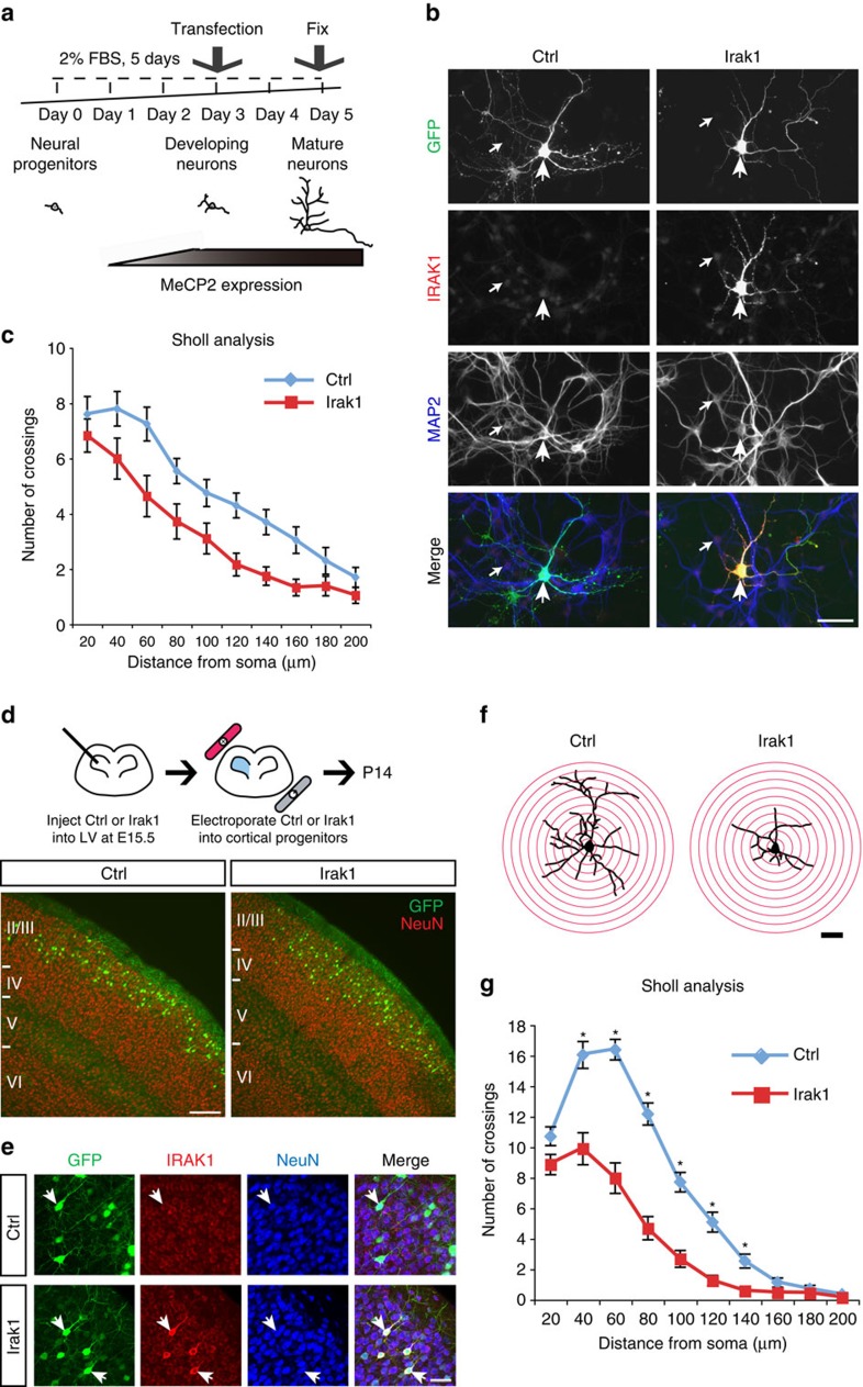
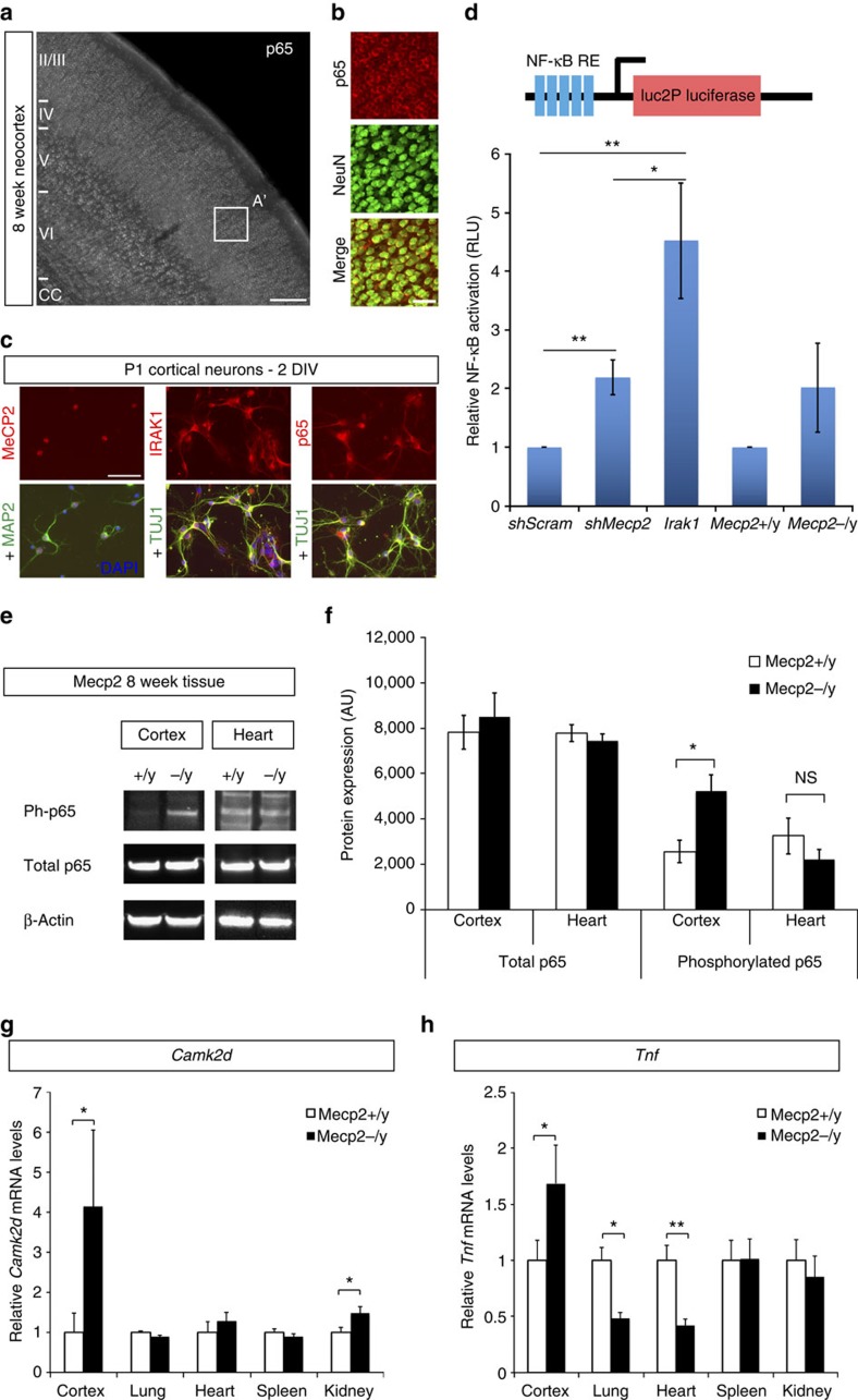
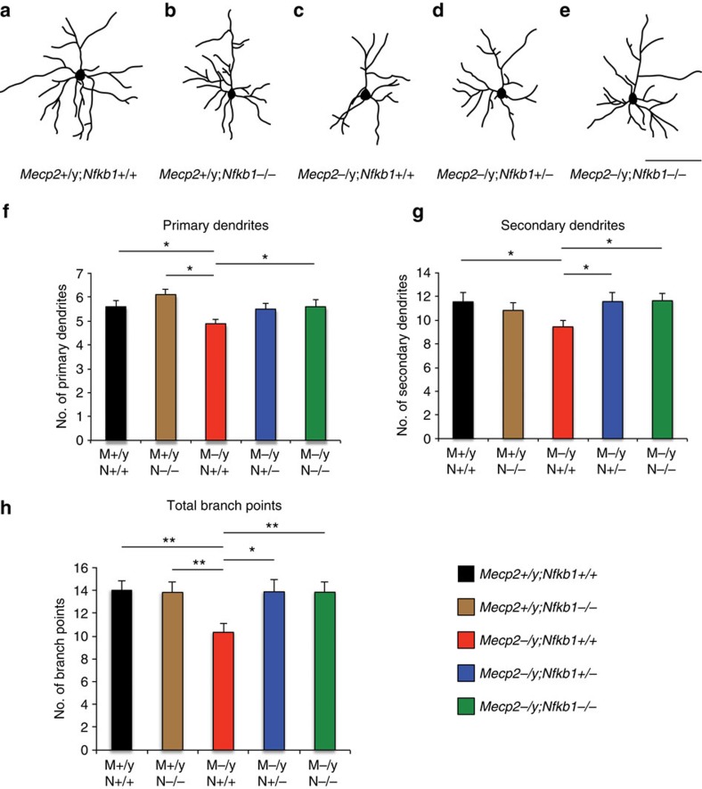
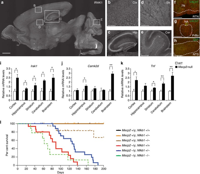
Mutations in the transcriptional regulator Mecp2 cause the severe X-linked neurodevelopmental disorder Rett syndrome (RTT). In this study, we investigate genes that function downstream of MeCP2 in cerebral cortex circuitry, and identify upregulation of Irak1, a central component of the NF-κB pathway. We show that overexpression of Irak1 mimics the reduced dendritic complexity of Mecp2-null cortical callosal projection neurons (CPN), and that NF-κB signalling is upregulated in the cortex with Mecp2 loss-of-function. Strikingly, we find that genetically reducing NF-κB signalling in Mecp2-null mice not only ameliorates CPN dendritic complexity but also substantially extends their normally shortened lifespan, indicating broader roles for NF-κB signalling in RTT pathogenesis. These results provide new insight into both the fundamental neurobiology of RTT, and potential therapeutic strategies via NF-κB pathway modulation.


Figures

Figure 1
Figure 1. MeCP2 represses Irak1 expression in a brain-specific manner.
(a) In our microarray analyses, three independent probe sets reveal that Irak1 is overexpressed an average of 2.9-fold in P14 Mecp2-null CPN, which was confirmed by qPCR on three independent biological replicates. *P<0.05, **P<0.01, paired two-sided t-test. (b) qPCR at 6 weeks of age reveals that Irak1 is overexpressed in cortices of two independent Mecp2-null mouse lines (B-mice and J-mice), when compared with wild-type cortex, but not in multiple non-CNS tissues. *P<0.05, unpaired two-sided t-test, mean±s.e.m. (cf) To exclude the possibility that overexpression of Irak1 is an artefact of gene targeting of Mecp2, we knocked down Mecp2 in wild-type layer 2/3 neurons by in utero electroporation of an Mecp2 shRNA, in comparison with a control shRNA, at E15.5. (c) Cells electroporated with Mecp2 shRNA do not exhibit any disruptions in migration, laminar location or survival in comparison to control, scrambled shRNA, as indicated by GFP reporter expression. Scale bar, 200 μm. (d) Electroporated cells were purified by FACS at P14, and qPCR analysis reveals that Irak1 is overexpressed an average of 1.9-fold following an approximate 40% reduction in Mecp2 mRNA. *P<0.05 from three independent experiments, paired two-sided t-test. (e) MeCP2 protein (red) is detected in P14 layer 2/3 neurons (NeuN, blue) electroporated with a control, scrambled shRNA, but it is not detected in layer 2/3 neurons electroporated with an Mecp2 shRNA construct. (f) IRAK1 protein (red) is detected in layer 2/3 NeuN positive (blue) neurons electroporated with either scrambled or Mecp2 shRNA. Scale bar, 25 μm (e,f).
Figure 2
Figure 2. Overexpression of Irak1 in cortical neurons reduces dendritic arborization.
(a) A plasmid overexpressing either a reporter GFP (Ctrl) or both Irak1 and GFP (Irak1) was transfected into developing immature neurons in culture. (b) IRAK1 protein (red) is highly expressed by neurons (MAP2 positive; blue) transfected with Irak1 and GFP (green), while neurons transfected with GFP alone express only endogenous, low levels of IRAK1. Large arrowheads indicate transfected neurons; small arrows indicate neighbouring, untransfected neurons. Scale bar, 50 μm. (c) Cells were immuno-labelled against both MAP2 and GFP, and the dendritic morphology of MAP2+/GFP+ cells was analysed by Sholl analysis. Overexpression of Irak1 significantly decreases neuronal dendritic arborization, compared to that of control neurons (control n=20, Irak1-overexpressing neurons n=36); two-way ANOVA, F(1,540)=39.9, P<0.001). Error bars=mean±s.e.m. (d) To investigate whether overexpression of Irak1 also modifies dendritic complexity of layer 2/3 neurons in vivo, Ctrl and Irak1 plasmids were injected in utero into the lateral ventricle (LV), and electroporated into neural precursors in the ventricular zone (VZ) at E15.5. Electroporated neural precursors subsequently give rise to differentiated cortical layer 2/3 projection neurons (NeuN+, red), with no obvious disruption in laminar location or survival at P14 with Irak1 overexpression. Scale bar, 200 μm. (e) IRAK1 protein (red) is highly overexpressed by P14 layer 2/3 neurons electroporated with Irak1, but not Ctrl, compared to endogenous IRAK1 expression within cortical layer 2/3 at P14. Scale bar, 50 μm. (f,g) We analysed the dendritic morphology of electroporated layer 2/3 projection neurons at P14 by Sholl analysis (ctrl n=24, Irak1 n=28). In vivo overexpression of Irak1 in layer 2/3 projection neurons results in reduced dendritic arborization, recapitulating the dendritic phenotype in layer 2/3 projection neurons in Mecp2-null mice. *P<0.01, a two-way ANOVA and the Bonferroni test, mean±s.e.m., Scale bars, 50 μm.
Figure 3
Figure 3. NF-κB signalling is upregulated by cortical neurons with Mecp2 loss-of-function.
(a,b) The NF-κB subunit p65 (also known as RelA; grey in a, red in b) is widely expressed by neurons throughout the neocortex (NeuN positive, green), including CPN in layer 2/3. Scale bar, 200 μm. (c,d) We directly investigated whether MeCP2 and IRAK1 regulate NF-κB signalling in cortical neurons. (c) We confirmed that wild-type P1 neurons express MeCP2, IRAK1 and p65/RelA (all red), with a primarily cytoplasmic (inactive) localization at 2 days in vitro (DIV), as well as the neuronal markers MAP2 and TUJ1 (green). Scale bar, 50 μm. (d) We nucleofected P1 cortical neurons with an NF-κB luciferase reporter construct, and either a control shRNA construct, an Mecp2 shRNA construct, or an Irak1 expression construct, and measured NF-κB activity by luciferase assay at 2 DIV (n=5 independent experiments). Knockdown of Mecp2 results in an approximate 2.2-fold increase in activation of NF-κB over control, while overexpression of Irak1 increases NF-κB activation 4.5-fold on average. We additionally compared NF-κB luciferase reporter activity in P1 Mecp2 wild-type and Mecp2-null cortical neurons, and identified a trend towards increased NF-κB activation in Mecp2-nulls (n=3 independent experiments). RLU, relative luminescence units, normalized to a control luciferase construct. *P<0.05, **P<0.01, unpaired two-sided t-test, mean±s.e.m. (e,f) We investigated NF-κB activation in vivo in 8 week neocortex and heart by western blot for phosphorylated p65/RelA, relative to total p65/RelA, and (e) identified an approximately 2-fold increase in phosphorylated p65/RelA specifically in Mecp2−/y cortex compared with littermate Mecp2+/y (n=3 littermate pairs). AU, arbitrary units. *P<0.05, unpaired two-sided t-test, mean±s.e.m. (g,h) Further, qPCR experiments using mRNA from wild-type and Mecp2-null mice (four littermate pairs) at 8 weeks of age reveal that two downstream genes involved in NF-κB signalling, Camk2d (g) and Tnf (h), are upregulated in cortex, but are not widely upregulated in non-CNS tissues. Gapdh expression was used as an internal control. *P<0.05, **P<0.001, unpaired two-sided t-test, mean±s.e.m.
Figure 4
Figure 4. Reducing NF-κB signalling improves reduced dendritic complexity of Mecp2-null CPN.
To determine whether rough correction of abnormally activated NF-κB signalling due to overexpression of Irak1 can partially rescue the reduced dendritic arborization in Mecp2-null cortex, we generated Mecp2 and Nfkb1 double mutant mice, and visualized neuronal morphology by Golgi staining. Representative examples of camera lucida drawings of layer 2/3 pyramidal neurons in Mecp2+/y;Nfkb1+/+ (a, n=30); Mecp2+/y; Nfkb1−/− (b, n=18); Mecp2−/y;Nfkb1+/+ (c, n=18), Mecp2−/y;Nfkb1+/− (d, n=26); and Mecp2−/y; and Nfkb1−/− (e, n=20) mice at 8 weeks of age. The numbers of primary (f) and secondary dendrites (g), and total branch points (h) in Mecp2−/y;Nfkb1+/− and Mecp2−/y;Nfkb1−/− neurons are rescued (increased) compared with those of Mecp2−/y;Nfkb1+/+ neurons. *P<0.05, **P<0.01 unpaired two-sided t-test, mean±s.e.m. Scale bar, 100 μm (ad).
Figure 5
Figure 5. Reducing aberrant NF-κB signalling improves lifespan of Mecp2-null mice.
(a) IRAK protein is expressed by neurons widely throughout the P14 brain, including in the (b) neocortex, (c) hippocampus, (d) striatum and (e) cerebellum. (f,h) IRAK1 (red) is also expressed in many brainstem regions that are critical for respiratory function, including (F) the retrotrapezoidal nucleus (RTN), located ventral to the VAChT-positive (green) facial nucleus (7N); (g) the pre-Botzinger complex (PrBo), located ventral to the VAChT-positive (green) nucleus ambiguus; and (h) the somatostatin (SST; green) positive nucleus of the solitary tract (NTS). Scale bar, 1 mm (a); scale bar, 200 μm (bh). (ik) RNA was isolated from cortex, hippocampus, striatum, cerebellum and brainstem of P14 wild-type (n=3) and Mecp2-null mice (n=3). Expression levels of Irak1 (i) and downstream genes involved in NF-κB signalling, Camk2d (j) and Tnf (k), were analysed by qPCR, and normalized to the expression level of Gapdh. These genes are widely dysregulated in multiple brain regions, including the cortex and the brainstem (implicated in respiratory dysfunction in RTT). *P<0.05, **P<0.01, unpaired two-sided t-test, mean±s.e.m. (l) Kaplan–Meier survival curves for Mecp2+/y; Nfkb1+/+ (black line, n=21), Mecp2+/y; Nfkb1+/− (orange line, n=28), Mecp2+/y; Nfkb1−/− (brown line, n=19), Mecp2−/y; Nfkb1+/+ (red line, n=15), Mecp2−/y; Nfkb1+/− (blue line, n=21); and Mecp2−/y; Nfkb1−/− (green line, n=9). Log-rank survival analysis reveals that Mecp2−/y; Nfkb1+/− mice survive significantly and substantially longer to blinded standardized humane morbidity criteria (∼50% increase) than Mecp2−/y; Nfkb1+/+ mice (P=0.002; median survival 131 versus 89 days, respectively).

References

    1. Chahrour M. & Zoghbi H. Y. The story of Rett syndrome: from clinic to neurobiology. Neuron 56, 422–437 (2007) . - PubMed
    1. Amir R. E. et al. Rett syndrome is caused by mutations in X-linked MECP2, encoding methyl-CpG-binding protein 2. Nat. Genet. 23, 185–188 (1999) . - PubMed
    1. Gonzales M. L. & LaSalle J. M. The role of MeCP2 in brain development and neurodevelopmental disorders. Curr. Psychiatry Rep. 12, 127–134 (2010) . - PMC - PubMed
    1. Chen R. Z., Akbarian S., Tudor M. & Jaenisch R. Deficiency of methyl-CpG binding protein-2 in CNS neurons results in a Rett-like phenotype in mice. Nat. Genet. 27, 327–331 (2001) . - PubMed
    1. Guy J., Hendrich B., Holmes M., Martin J. E. & Bird A. A mouse Mecp2-null mutation causes neurological symptoms that mimic Rett syndrome. Nat. Genet. 27, 322–326 (2001) . - PubMed

Publication types

MeSH terms