

Ultrastructural comparison of porcine putative embryonic stem cells derived by in vitro fertilization and somatic cell nuclear transfer
- PMID: 26821870
- PMCID: PMC4848575
- DOI: 10.1262/jrd.2015-124
Ultrastructural comparison of porcine putative embryonic stem cells derived by in vitro fertilization and somatic cell nuclear transfer
Abstract
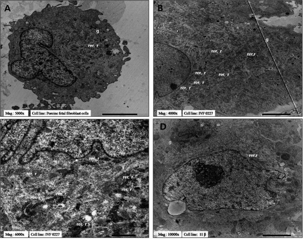
The ultrastructure of porcine putative embryonic stem cells and porcine fetal fibroblasts (PFFs) was analyzed by transmission electron microscopy. The aim of this study was to compare the features of organelles in in vitro fertilization (IVF) derived porcine embryonic stem cells (IVF-pESCs) and somatic cell nuclear transfer (SCNT) derived pESCs (SCNT-pESCs). Also, the features of organelles in high-passage IVF-pESCs were compared with those in low-passage cells. The ultrastructure of PFFs showed rare microvilli on the cell surfaces, polygonal or irregular nuclei with one to two reticular-shaped nucleoli and euchromatin, low cytoplasm-to-nucleus ratios, rare ribosomes, rare rough endoplasmic reticulum, elongated mitochondria, rich lysosomes and rich phagocytic vacuoles. IVF-pESCs showed rare microvilli on the cell surfaces, round or irregular nuclei with one to two reticular-shaped nucleoli and euchromatin, low cytoplasm-to-nucleus ratios, rich ribosomes, long stacks of rough endoplasmic reticulum, elongated mitochondria, rare lysosomes and rare autophagic vacuoles. By contrast, SCNT-pESCs showed rich microvilli with various lengths and frequencies on the cell surfaces, polygonal nuclei with one reticular shaped nucleoli and heterochromatin, high cytoplasm-to-nucleus ratios, rare ribosomes, rare rough endoplasmic reticulum, round mitochondria, rich lysosomes and rich phagocytic vacuoles with clear intercellular junctions. Furthermore, high-passage IVF-pESCs showed irregularly shaped colonies, pyknosis and numerous lysosomes associated with autophagic vacuoles showing signs of apoptosis. In conclusion, this study confirms that the ultrastructural characteristics of pESCs differ depending on their origin. These ultrastructural characteristics might be useful in biomedical research using pESCs, leading to new insights regarding regenerative medicine and tissue repair.
Figures


Similar articles
-
Ultrastructural characteristics of human granulosa cells in a coculture system for in vitro fertilization.Microsc Res Tech. 2006 Jun;69(6):508-16. doi: 10.1002/jemt.20309. Microsc Res Tech. 2006. PMID: 16718668
-
Ultrastructural changes in goat interspecies and intraspecies reconstructed early embryos.Zygote. 2008 May;16(2):93-110. doi: 10.1017/S0967199407004492. Zygote. 2008. PMID: 18405430
-
Ultrastructural characteristics of three undifferentiated mouse embryonic stem cell lines and their differentiated three-dimensional derivatives: a comparative study.Cell Reprogram. 2014 Apr;16(2):151-65. doi: 10.1089/cell.2013.0073. Epub 2014 Mar 7. Cell Reprogram. 2014. PMID: 24606239 Free PMC article.
-
From oogonia to mature oocytes: inactivation of the maternal centrosome in humans.Microsc Res Tech. 2006 Jun;69(6):396-407. doi: 10.1002/jemt.20299. Microsc Res Tech. 2006. PMID: 16718650 Review.
-
Somatic cell nuclear transfer and derivation of embryonic stem cells in the mouse.Methods. 2008 Jun;45(2):101-14. doi: 10.1016/j.ymeth.2008.04.002. Epub 2008 Jun 2. Methods. 2008. PMID: 18593608 Review.
Cited by
-
Apoptosis in Porcine Pluripotent Cells: From ICM to iPSCs.Int J Mol Sci. 2016 Sep 12;17(9):1533. doi: 10.3390/ijms17091533. Int J Mol Sci. 2016. PMID: 27626414 Free PMC article. Review.
-
Diverse autophagy and apoptosis in myeloid leukemia cells induced by 20(s)-GRh2 and blue LED irradiation.RSC Adv. 2019 Nov 28;9(67):39124-39132. doi: 10.1039/c9ra08049j. eCollection 2019 Nov 27. RSC Adv. 2019. PMID: 35540666 Free PMC article.
-
Blastocyst-like Structures in the Peripheral Retina of Young Adult Beagles.Int J Mol Sci. 2024 May 30;25(11):6045. doi: 10.3390/ijms25116045. Int J Mol Sci. 2024. PMID: 38892233 Free PMC article.
References
-
- Thomson JA, Marshall VS. Primate embryonic stem cells. Curr Top Dev Biol 1998; 38: 133–165. - PubMed
-
- Thomson JA, Itskovitz-Eldor J, Shapiro SS, Waknitz MA, Swiergiel JJ, Marshall VS, Jones JM. Embryonic stem cell lines derived from human blastocysts. Science 1998; 282: 1145–1147. - PubMed
-
- Takahashi K, Yamanaka S. Induction of pluripotent stem cells from mouse embryonic and adult fibroblast cultures by defined factors. Cell 2006; 126: 663–676. - PubMed
-
- Friel R, van der Sar S, Mee PJ. Embryonic stem cells: understanding their history, cell biology and signalling. Adv Drug Deliv Rev 2005; 57: 1894–1903. - PubMed
-
- Wobus AM. Potential of embryonic stem cells. Mol Aspects Med 2001; 22: 149–164. - PubMed
MeSH terms
LinkOut - more resources
Full Text Sources
Other Literature Sources