. 2016 Jan 29:16:13.
doi: 10.1186/s12876-016-0423-6.

Development of a transgenic mouse model of hepatocellular carcinoma with a liver fibrosis background

Affiliations

Development of a transgenic mouse model of hepatocellular carcinoma with a liver fibrosis background

Sook In Chung et al. BMC Gastroenterol. .

Abstract

Background: Liver fibrosis and its end-stage disease, cirrhosis, are major risk factors for hepatocellular carcinoma (HCC) and present in 80 to 90 % of patients with HCC. Current genetically engineered mouse models for HCC, however, generally do not feature liver fibrosis, which is a critical discrepancy between human HCC and murine models thereof. In this study, we developed a simple transgenic mouse model of HCC within the context of a fibrotic liver.

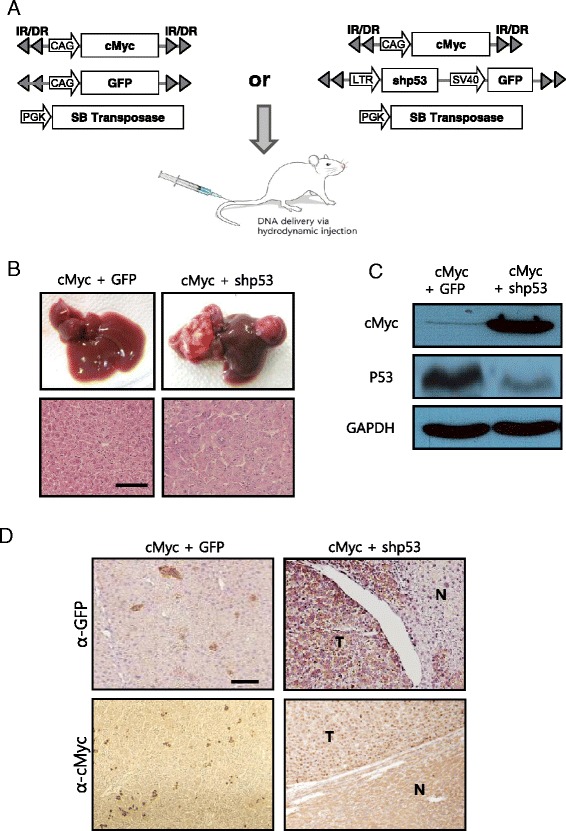
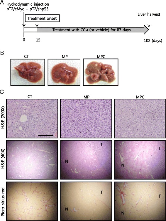
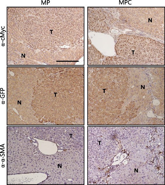
Methods: Employing hydrodynamic transfection (HT), coupled with the Sleeping Beauty (SB) transposon system, liver was stably transfected with transposons expressing cMyc and a short hairpin RNA down-regulating p53 (shp53). A chronic liver injury model, induced by hepatotoxic carbon tetrachloride (CCl4), was applied to the transgenic mice, allowing cells expressing cMyc plus shp53 to become malignant in the background of liver fibrosis.

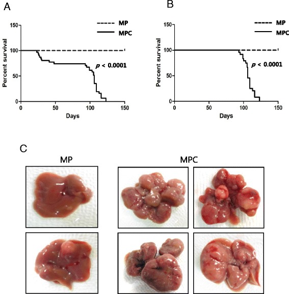
Results: Livers harvested about 3 months after HT had excessive collagen deposition and activated hepatic stellate cells surrounding the tumors. Hepatocarcinogenesis was significantly accelerated in the fibrotic livers compared to those of the control, significantly decreasing the life span of the mice. The tumor incidence and average number of tumors per mouse were significantly higher in the group treated with CCl4 compared to the vehicle-treated control mice, following HT (p < 0.01).

Conclusions: Considering the simplicity and efficiency in generating HCC for fibrotic livers, the transgenic HCC model has the potential to be effectively used in preclinical testing of HCC anticancer therapy and in studies of hepatocarcinogenesis in fibrotic livers.


Figures

Fig. 1
Fig. 1
Expression of cMyc plus shp53 in the liver induces well-differentiated HCC. a Schematic illustration of the experimental procedure to generate transgenic livers expressing cMyc plus GFP (control), and cMyc plus shp53. Hydrodynamic transfection was performed using a mixture of indicated plasmids. b Gross morphology (upper panels) of livers harvested from mice of each group at 7 months post hydrodynamic injection. Images of H&E staining in liver sections are shown below. Well-differentiated HCCs developed in cMyc plus shp53 mice, while the control mice did not develop tumors. Scale bar, 100 μm. c Protein expression levels of cMyc and p53 in liver of cMyc plus GFP mice (control) and liver tumor of cMyc plus shp53 mice. d Images of IHC staining for GFP and cMyc in liver sections of indicated groups. “T” denotes tumor and “N” denotes non-tumorous liver parenchyma tissue. Scale bar, 100 μm
Fig. 2
Fig. 2
Application of a chronic liver injury model to the cMyc plus shp53 mice induces HCC in a fibrotic liver background. a Diagram of experimental procedures. Starting at 15 d post hydrodynamic transfection, mice were treated with CCl4 or the vehicle twice per week for about 12 weeks. b Gross images of representative livers of CT, MP, and MPC mice that were harvested at 102 d post hydrodynamic transfection. c H&E (200× and 40×) and picro-sirius red staining in liver sections of indicated groups. Tumors from both MP and MPC show similar well-differentiated HCC phenotypes (upper panels). Low-magnification images of the tumor seen in upper panels reveal boundaries between tumor (T) areas and areas of non-tumorous (N) liver parenchyma tissue (middle panels). Images of picro-sirius red staining from the same area seen in middle panels are presented in lower panels. Note the presence of accumulated collagen bands in the area surrounding the liver tumor in an MPC mouse. Scale bars, 200 μm for upper panels and 1 mm for middle and lower panels
Fig. 3
Fig. 3
Images of IHC staining for cMyc, GFP and α-SMA in tumor sections. Tumors from both MP and MPC mice were stained positive for cMyc and GFP. Note that cMyc was localized in the nuclei of tumor cells. The area surrounding tumor of MPC mice shows the presence of activated hepatic stellate cells, based on α-SMA staining. “T” denotes tumor and “N” denotes non-tumorous liver parenchyma tissue. Scale bar, 200 μm
Fig. 4
Fig. 4
Short life span of MPC mice and increased tumor burdens in their livers. a, b Kaplan–Meier survival curves of MP and MPC mice in the entire cohort (a) and with the exclusion of mice that died initially due to CCl4-induced toxicity (b). Note that MPC mice had a significantly shorter life span compared to MP mice, even with the exclusion of the mice that died early in the MPC group (p < 0.0001). c Gross images of representative livers of MP and MPC mice. Livers were harvested following death for the MPC group and at the end of the experiment in the MP group

References

    1. Ince N, Wands JR. The increasing incidence of hepatocellular carcinoma. N Engl J Med. 1999;340:798–9. doi: 10.1056/NEJM199903113401009. - DOI - PubMed
    1. El-Serag HB. Hepatocellular carcinoma. N Engl J Med. 2011;365:1118–27. doi: 10.1056/NEJMra1001683. - DOI - PubMed
    1. Hassan MM, Hwang LY, Hatten CJ, Swaim M, Li D, Abbruzzese JL, et al. Risk factors for hepatocellular carcinoma: synergism of alcohol with viral hepatitis and diabetes mellitus. Hepatology. 2002;36:1206–13. doi: 10.1053/jhep.2002.36780. - DOI - PubMed
    1. El-Serag HB, Rudolph KL. Hepatocellular carcinoma: epidemiology and molecular carcinogenesis. Gastroenterology. 2007;132:2557–76. doi: 10.1053/j.gastro.2007.04.061. - DOI - PubMed
    1. Ascha MS, Hanouneh IA, Lopez R, Tamimi TA, Feldstein AF, Zein NN. The incidence and risk factors of hepatocellular carcinoma in patients with nonalcoholic steatohepatitis. Hepatology. 2010;51:1972–8. doi: 10.1002/hep.23527. - DOI - PubMed

Publication types

MeSH terms