

Constructal approach to cell membranes transport: Amending the 'Norton-Simon' hypothesis for cancer treatment
- PMID: 26822208
- PMCID: PMC4731791
- DOI: 10.1038/srep19451
Constructal approach to cell membranes transport: Amending the 'Norton-Simon' hypothesis for cancer treatment
Abstract
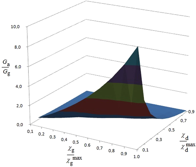
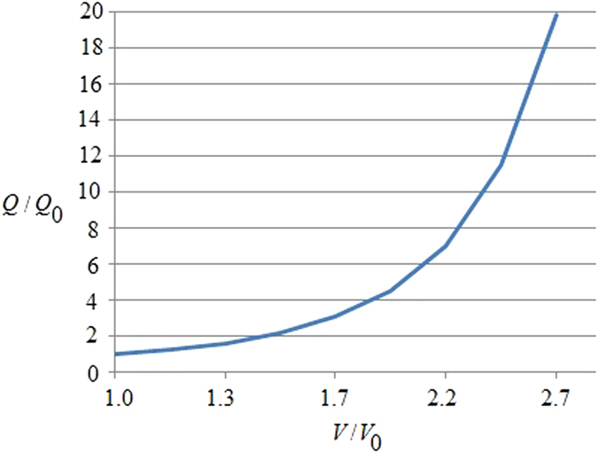
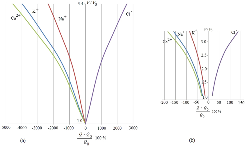
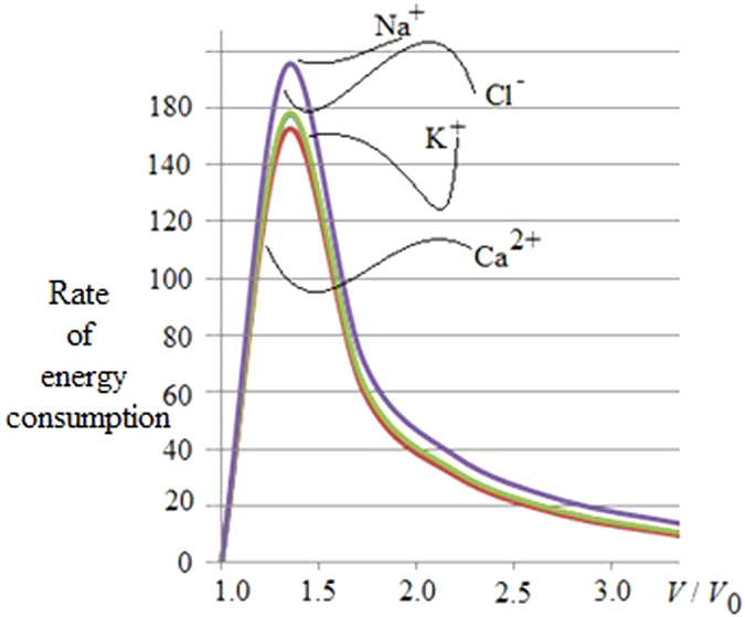
To investigate biosystems, we propose a new thermodynamic concept that analyses ion, mass and energy flows across the cell membrane. This paradigm-shifting approach has a wide applicability to medically relevant topics including advancing cancer treatment. To support this claim, we revisit 'Norton-Simon' and evolving it from an already important anti-cancer hypothesis to a thermodynamic theorem in medicine. We confirm that an increase in proliferation and a reduction in apoptosis trigger a maximum of ATP consumption by the tumor cell. Moreover, we find that positive, membrane-crossing ions lead to a decrease in the energy used by the tumor, supporting the notion of their growth inhibitory effect while negative ions apparently increase the cancer's consumption of energy hence reflecting a growth promoting impact. Our results not only represent a thermodynamic proof of the original Norton-Simon hypothesis but, more concretely, they also advance the clinically intriguing and experimentally testable, diagnostic hypothesis that observing an increase in negative ions inside a cell in vitro, and inside a diseased tissue in vivo, may indicate growth or recurrence of a tumor. We conclude with providing theoretical evidence that applying electromagnetic field therapy early on in the treatment cycle may maximize its anti-cancer efficacy.
Figures
Similar articles
-
The importance of ion fluxes for cancer proliferation and metastasis: A thermodynamic analysis.J Theor Biol. 2018 May 14;445:1-8. doi: 10.1016/j.jtbi.2018.02.019. Epub 2018 Feb 21. J Theor Biol. 2018. PMID: 29474857
-
Model of active transport of ions in biomembranes based on ATP-dependent change of height of diffusion barriers to ions.J Theor Biol. 2006 Oct 7;242(3):617-26. doi: 10.1016/j.jtbi.2006.04.011. Epub 2006 Apr 28. J Theor Biol. 2006. PMID: 16750835
-
Requirements on models and models of active transport of ions in biomembranes.Bull Math Biol. 2006 Feb;68(2):385-99. doi: 10.1007/s11538-005-9035-y. Epub 2006 Mar 30. Bull Math Biol. 2006. PMID: 16794936
-
[Possible role of cholesterol in cellular membranes].Usp Sovrem Biol. 1974 May-Jun;77(3):331-47. Usp Sovrem Biol. 1974. PMID: 4214364 Review. Russian. No abstract available.
-
Overview: mechanism of free energy coupling between ATP hydrolysis and ion transport.Prog Clin Biol Res. 1988;268A:401-20. Prog Clin Biol Res. 1988. PMID: 2971220 Review. No abstract available.
Cited by
-
Magnetic field potential effects on the doxorubicin therapeutic activity in Ehrlich tumor growth.Saudi J Biol Sci. 2021 Apr;28(4):2566-2574. doi: 10.1016/j.sjbs.2021.01.061. Epub 2021 Feb 11. Saudi J Biol Sci. 2021. PMID: 33935572 Free PMC article.
-
Rate of entropy model for irreversible processes in living systems.Sci Rep. 2017 Aug 22;7(1):9134. doi: 10.1038/s41598-017-09530-5. Sci Rep. 2017. PMID: 28831153 Free PMC article.
-
A Thermodynamic Perspective of Cancer Cells' Volume/Area Expansion Ratio.Membranes (Basel). 2023 Nov 30;13(12):895. doi: 10.3390/membranes13120895. Membranes (Basel). 2023. PMID: 38132898 Free PMC article.
-
Predicting cancer stages from tissue energy dissipation.Sci Rep. 2023 Sep 23;13(1):15894. doi: 10.1038/s41598-023-42780-0. Sci Rep. 2023. PMID: 37741864 Free PMC article.
References
-
- Pelling A. E., Sehati S., Gralla E. B., Valentine J. S. & Gimzewski J. K. Local nanomechanical motion of the cell wall of Saccharomyces cerevisiae. Science 305, 1147–1150 (2004). - PubMed
-
- Venkatasubramanian R., Henson M. A. & Forbes N. S. Incorporating energy metabolism into a growth model of multicellular tumor spheroids. J. Theor. Biol. 242, 440–453 (2006). - PubMed
Publication types
MeSH terms
Substances
LinkOut - more resources
Full Text Sources
Other Literature Sources
