. 2017 Jan;27(1):64-76.
doi: 10.1111/bpa.12362. Epub 2016 May 6.

SLC20A2 Deficiency in Mice Leads to Elevated Phosphate Levels in Cerbrospinal Fluid and Glymphatic Pathway-Associated Arteriolar Calcification, and Recapitulates Human Idiopathic Basal Ganglia Calcification

Affiliations

SLC20A2 Deficiency in Mice Leads to Elevated Phosphate Levels in Cerbrospinal Fluid and Glymphatic Pathway-Associated Arteriolar Calcification, and Recapitulates Human Idiopathic Basal Ganglia Calcification

Mary Catherine Wallingford et al. Brain Pathol. 2017 Jan.

Abstract

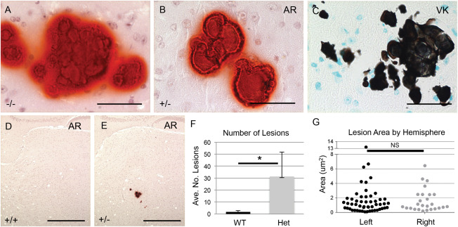
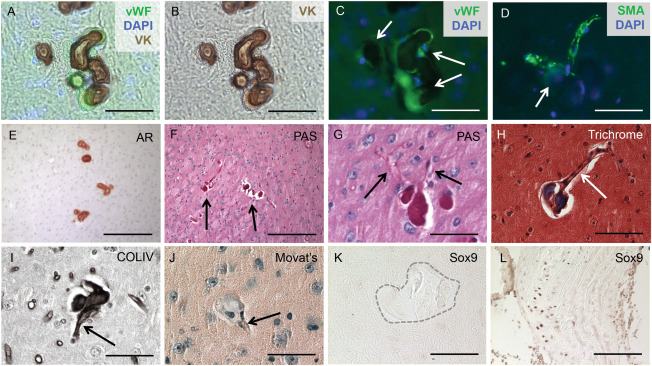
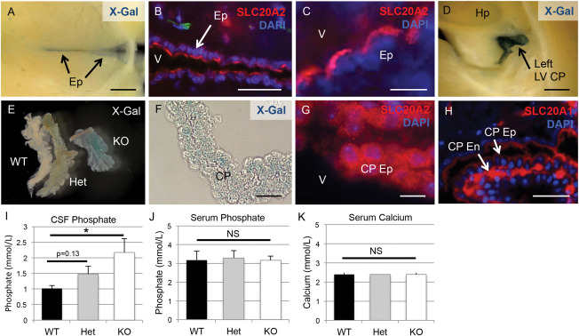
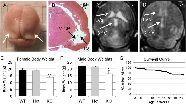
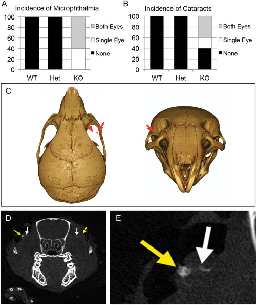
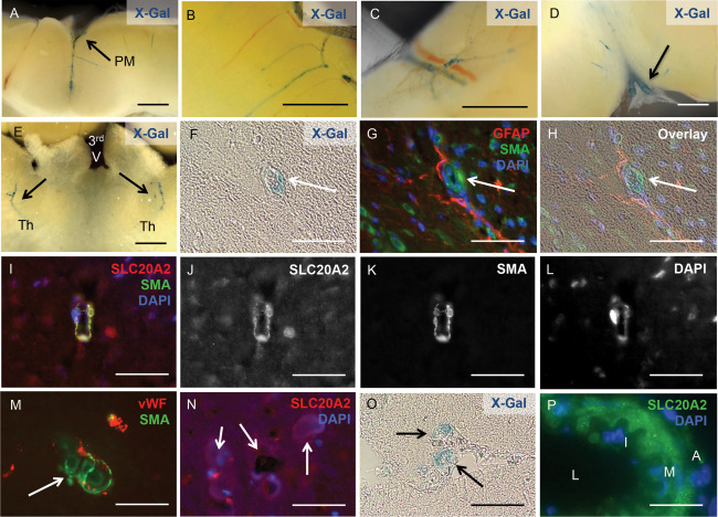
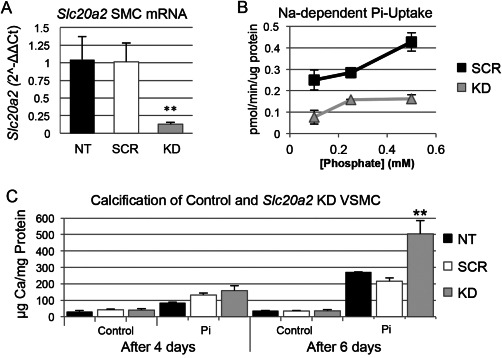
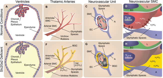
Idiopathic basal ganglia calcification is a brain calcification disorder that has been genetically linked to autosomal dominant mutations in the sodium-dependent phosphate co-transporter, SLC20A2. The mechanisms whereby deficiency of Slc20a2 leads to basal ganglion calcification are unknown. In the mouse brain, we found that Slc20a2 was expressed in tissues that produce and/or regulate cerebrospinal fluid, including choroid plexus, ependyma and arteriolar smooth muscle cells. Haploinsufficient Slc20a2 +/- mice developed age-dependent basal ganglia calcification that formed in glymphatic pathway-associated arterioles. Slc20a2 deficiency uncovered phosphate homeostasis dysregulation characterized by abnormally high cerebrospinal fluid phosphate levels and hydrocephalus, in addition to basal ganglia calcification. Slc20a2 siRNA knockdown in smooth muscle cells revealed increased susceptibility to high phosphate-induced calcification. These data suggested that loss of Slc20a2 led to dysregulated phosphate homeostasis and enhanced susceptibility of arteriolar smooth muscle cells to elevated phosphate-induced calcification. Together, dysregulated cerebrospinal fluid phosphate and enhanced smooth muscle cell susceptibility may predispose to glymphatic pathway-associated arteriolar calcification.

Keywords: Slc20a2; cerebral vascular calcification; cerebrospinal fluid; glymphatic spaces; idiopathic basal ganglia calcification; phosphate.


Figures

Figure 1
Figure 1
Slc20a2 deficient mice develop BGC. BGC presence was detected in Slc20a2 −/− mice by Alizarin Red (AR) (A). Brain sections from Slc20a2 +/− mice revealed mineral deposits positive for both AR (B) and von Kossa (VK) (C). Abundant calcified lesions were observed by AR staining in Slc20a2 +/− mice but not Slc20a2 +/+ controls (D, E). The average number of lesions was significantly higher in the Slc20a2 +/− mice compared to the Slc20a2 +/+ controls as determined by a two‐tailed Student's T‐test with unequal variance; N = 6; P‐value=0.018 (F). As seen in human BGC patients, the calcified lesions were located bilaterally as determined by a two‐tailed Wilcoxon signed ranks test; N = 6; P‐value=0.7265 (G). Scale bars: 50 μM (A‐C), 500 μM (D,E).
Figure 2
Figure 2
BGC is localized to cerebral arterioles. The calcified lesions (arrows in C) were localized to arterioles, characterized by the co‐localization of vWF and von Kossa (VK) staining and counterstaining with the nuclear marker DAPI (A‐C). Furthermore, the lesions (arrow in D) were also present in SMA‐positive vessels counterstained with DAPI (D). Alizarin Red (AR) staining compared with Periodic acid‐Schiff (PAS)‐positive staining of consecutive sections confirmed that the calcified lesions (arrows in F) contained basement membrane components (E‐G) and localized to blood vessels (arrows in G). Positive Masson's Trichrome staining indicated that the lesions were rich in collagen and/or mucin (H) and formed in blood vessels (arrow in Figure H). Immunohistochemical staining identified collagen type IV as the major basement membrane protein present in the calcified lesions (I) and further confirmed localization to blood vessels (arrow in I). The SMC in the calcified regions do not undergo an osteochondrogenic phenotype change, as the lesions (arrow in J) lacked proteoglycan‐rich or cartilage‐like structures, confirmed by negative Movat Pentachrome (J) and SOX9 (K) staining. Calcified aortic tissue from a diabetes mouse model was used as a positive control (L). Scale bars: 50μM (A‐D,G‐L), 200μM (E,F).
Figure 3
Figure 3
Slc20a2 is expressed in CSF‐associated tissues and regulates CSF phosphate levels. X‐Gal staining and/or immunofluorescence counterstain with the nuclear marker DAPI were used to determine tissue‐specific expression patterns of Slc20a2. Both X‐Gal staining and antibody mediated detection of revealed localization in neurovascular tissues involved in cerebrospinal fluid (CSF) regulation, including the ependyma (Ep) (A‐C) and the choroid plexus (CP) epithelium (Ep) (D‐G). Furthermore, immunohistochemical staining localized SLC20A1 to the choroid plexus endothelium (CP En) but not the CP Ep (H). CSF phosphate levels were trending in Slc20a2 +/− mice and significantly increased in the Slc20a2 −/− mice compared to the WT mice as determined by a One‐way ANOVA followed by Tukey's Post Hoc Test; N = 11; KO P‐value is < 0.05 when tested against WT samples (I). Data shown is representative of three independent experiments. Compared to Slc20a2 +/+ mice, serum phosphate and calcium levels in both the Slc20a2 +/− and Slc20a2 −/− mice were not significantly different, as determined by a One‐way ANOVA; N = 23 (J, K). Scale bars: 2 mm (A,D,E), 50μM (B), 10μM (C,F,G), 20μM (H). Abbreviations: CP En=choroid plexus endothelium; CP Ep=choroid plexus epithelium; Ep=ependyma; LV=lateral ventricle; V=ventricle.
Figure 4
Figure 4
Complete Slc20a2 loss results in hydrocephalus and premature death. Severe lethal hydrocephalus cases and premature death were seen in our weaned Slc20a2 −/− mice, as diagnosed by brain morphology (A) and by histology of H&E stained coronal sections (B). Moderate to severe hydrocephalus was confirmed in Slc20a2 −/− mice by MRI, n = 2 (C) and detection of ventricles in Slc20a2 +/− mice by MRI indicates mild hydrocephalus (n = 1) in Slc20a2 +/− mice (D). Reduced body weights were observed in both Slc20a2 −/− female (N = 25; P‐value<0.01) and Slc20a2 −/− male (N = 21; P‐value<0.05) mice when compared to age and sex matched Slc20a2 +/+ controls at six weeks of age, determined by a One‐way ANOVA followed by Tukey's Post Hoc Test (E,F). Poor survival was observed in Slc20a2 −/− mice between 4 and 21 weeks of age (G).
Figure 5
Figure 5
Ocular abnormalities associated with Slc20a2 deficiency. Both microphthalmia and cataracts were observed in six week old Slc20a2 KO mice (WT n = 8; Het n = 4; KO n = 5) (A,B). Furthermore, 9 μM CT scans detected calcification (red arrows in C) in both cataracts (yellow arrow in D,E) and in close proximity to the optic nerve head (white arrows in D,E).
Figure 6
Figure 6
Slc20a2 is highly expressed in cerebral SMC. X‐Gal activity was detected in arteries within the pia mater (PM) (A), the cortex (B), the cerebellum (C), in the region of the cervical lymphatics (D), and in the thalamus of both hemispheres (Th); location of the third ventricle (3rd V) is indicated for spatial orientation (E). X‐Gal staining and anti‐SLC20A2 antibody signal co‐localized with SMA‐positive cells and GFAP‐positive cell projections (F‐L). Calcified lesions (arrows in M, N) also co‐localized with vWF and SMA positive cells (M), and revealed the presence of SLC20A2 positive cells in low numbers (N). Slc20a2 was also expressed in media of vessels that connect to the CP (O) and in large cerebral arteries (L: lumen, I: intima, M: media, A: adventitia) (P). Scale bars: 2mm (A‐E), 50μM (F‐P).
Figure 7
Figure 7
Loss of Slc20a2 in SMC results in a susceptibility to high phosphate‐induced calcification. Knockdown (KD) of Slc20a2 in SMCs was induced by short interfering RNA (siRNA). The level of Slc20a2 in KD SMCs was 90% on average (P‐value<0.0001) compared to the NT and scramble siRNA treated (SCR) controls (n = 3 per condition), determined by a One‐way ANOVA with Tukey's Post Hoc Test (A). Furthermore, the KD SMCs (n = 3 per concentration) had a lower phosphate uptake at all phosphate concentrations compared to SCR controls (n = 3 per concentration): 0.1 mM phosphate (P‐value=0.0085); 0.25 mM phosphate (P‐value=0.011); and 0.5 mM phosphate (P‐value=0.0027) determined by a two‐tailed Student's T‐test with unequal variance (B). Cultured SMCs (n = 3 per condition) were also treated with normal or high phosphate media calcification media. Calcification was time‐dependent; after 6 days, the KD SMCs had an increased level of calcification (P‐value=0.006) compared to the NT and SCR controls, using a One‐way ANOVA with Tukey's Post Hoc Test (C).
Figure 8
Figure 8
Slc20a2 deficiency‐associated high CSF phosphate and SMC susceptibility to calcification may result in BGC in a 2‐hit mechanism. In normal conditions phosphate CSF concentrations are maintained at homeostatic levels as indicated in white in A‐D. SLC20A2 is poised to regulate CSF phosphate and is localized to the choroid plexus and ependyma (red in A), and the neurovascular SMCs (red in B‐D) adjacent to CSF‐containing glymphatic spaces. In Slc20a2 deficient conditions, these tissues contain reduced levels of SLC20A2 indicated in brown (E‐H). Slc20a2 deficiency leads to higher CSF phosphate concentrations in glymphatic spaces as indicated in yellow (E‐H). The glymphatic spaces are delineated by astrocytes and permeated by astrocyte endfeet, creating pockets. Fluid flow through this porous and segmented structure (G and H), is assumed to increase retention parameters caused by turbulent flow and increased surface‐ion interactions mediated by Brownian Forces. In this case a higher concentration of phosphate likely accumulates in glymphatic spaces that surround the exterior of smaller vessels, indicated by the yellow gradient and segmentation by astrocyte endfeet (F‐H). In normal conditions, SLC20A2 may act to promote SMC phosphate clearance from arteriolar glymphatic spaces (red arrows in D), and SLC20A2 plays a currently unidentified protective role against SMC calcification, illustrated by active gene expression in a SMC nucleus (D). In disease conditions, SLC20A2 deficiency results in high CSF phosphate concentrations compounded by the loss of protective roles against SMC calcification (H). We pose the hypothesis that these abnormalities lead to BGC in a 2‐hit mechanism. Abbreviations: BGC=basal ganglia calcification; SMC = smooth muscle cell; EC = endothelial cell; A = astrocyte; L = lumen.

Comment in

Similar articles

Cited by

References

    1. Betsholtz C, Keller A (2014) PDGF, pericytes and the pathogenesis of idiopathic basal ganglia calcification (IBGC). Brain Pathol 24:387–395. - PMC - PubMed
    1. Chen WJ et al (2013) Novel SLC20A2 mutations identified in southern Chinese patients with idiopathic basal ganglia calcification. Gene 529:159–162. - PubMed
    1. Cheng SL, Shao JS, Charlton‐Kachigian N, Loewy AP, Towler DA (2003) MSX2 promotes osteogenesis and suppresses adipogenic differentiation of multipotent mesenchymal progenitors. J Biol Chem 278:45969–45977. - PubMed
    1. Crouthamel MH et al (2013) Sodium‐dependent phosphate cotransporters and phosphate‐induced calcification of vascular smooth muscle cells: redundant roles for PiT‐1 and PiT‐2. Arteriosclerosis, Thrombosis, Vascular Biol 33:2625–2632. - PMC - PubMed
    1. Demoulin JB et al (2004) Platelet‐derived growth factor stimulates membrane lipid synthesis through activation of phosphatidylinositol 3‐kinase and sterol regulatory element‐binding proteins. J Biol Chem 279:35392–35402. - PubMed

MeSH terms

Substances

Supplementary concepts