. 2016 May 1;310(9):G705-15.
doi: 10.1152/ajpgi.00314.2015. Epub 2016 Jan 28.

Rapid disruption of intestinal epithelial tight junction and barrier dysfunction by ionizing radiation in mouse colon in vivo: protection by N-acetyl-l-cysteine

Affiliations

Rapid disruption of intestinal epithelial tight junction and barrier dysfunction by ionizing radiation in mouse colon in vivo: protection by N-acetyl-l-cysteine

Pradeep K Shukla et al. Am J Physiol Gastrointest Liver Physiol. .

Abstract

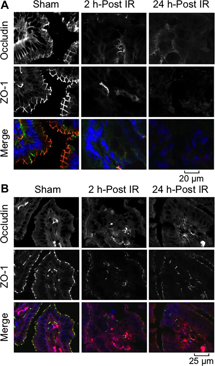
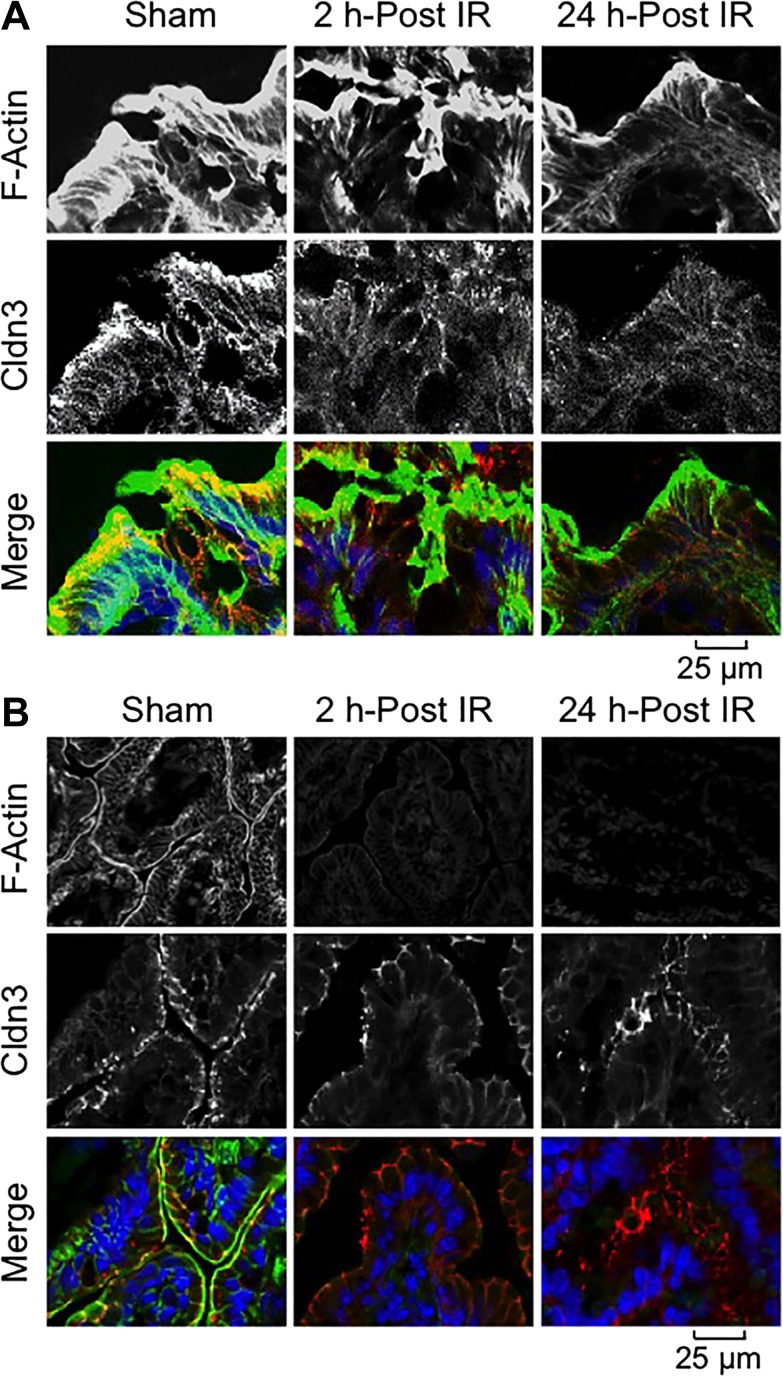
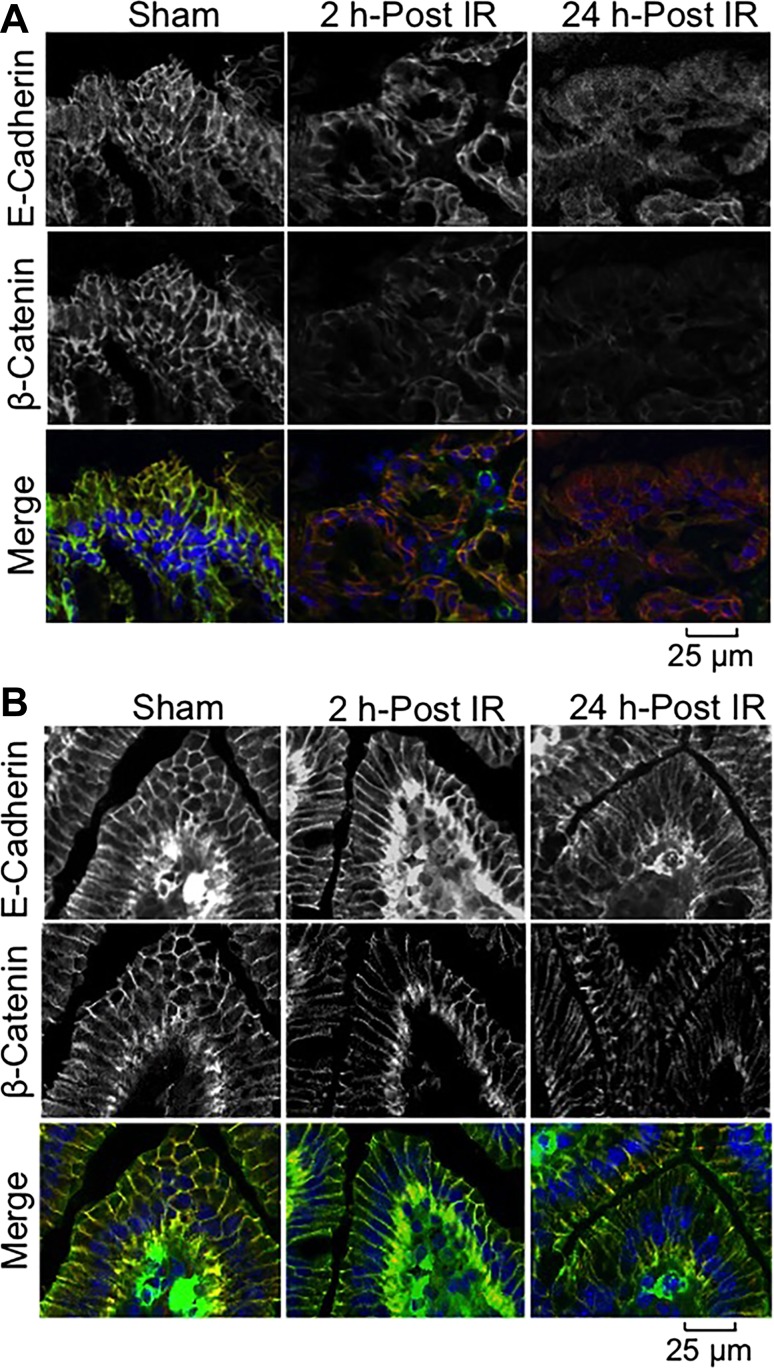
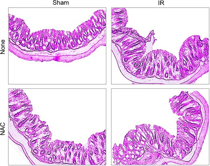
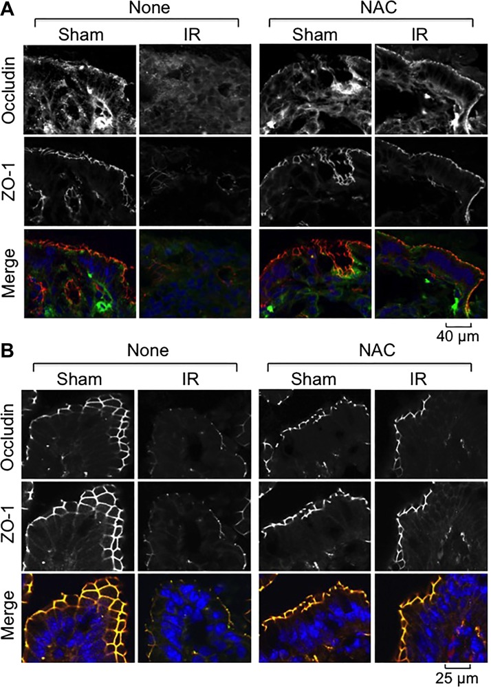
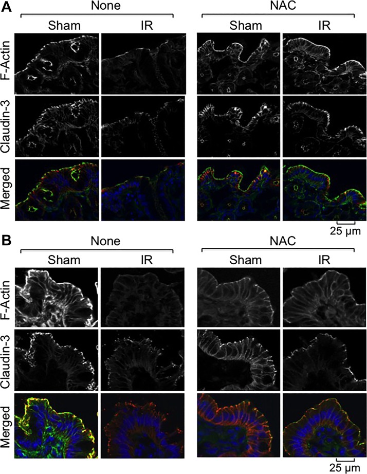
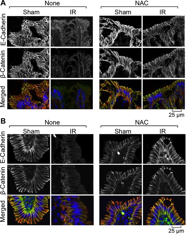
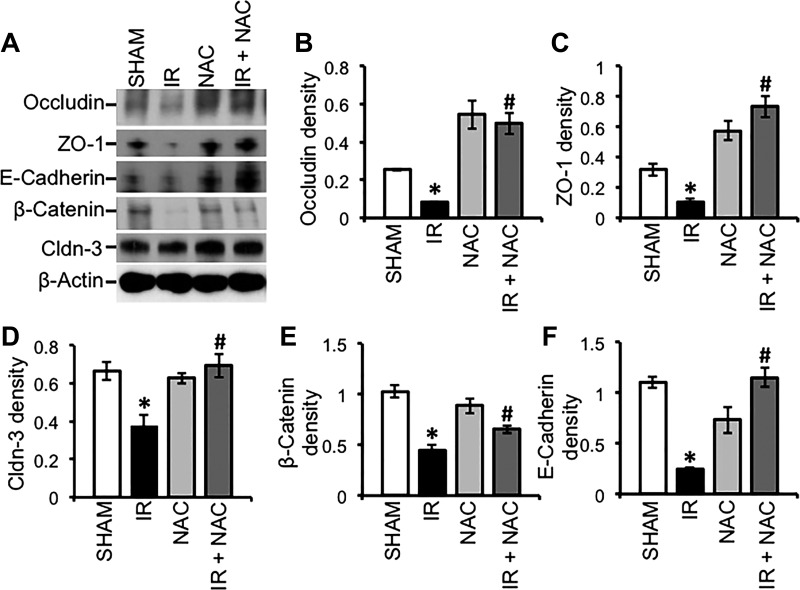
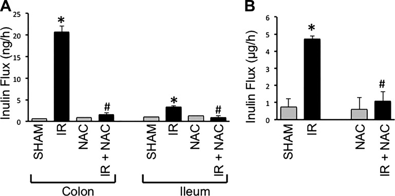
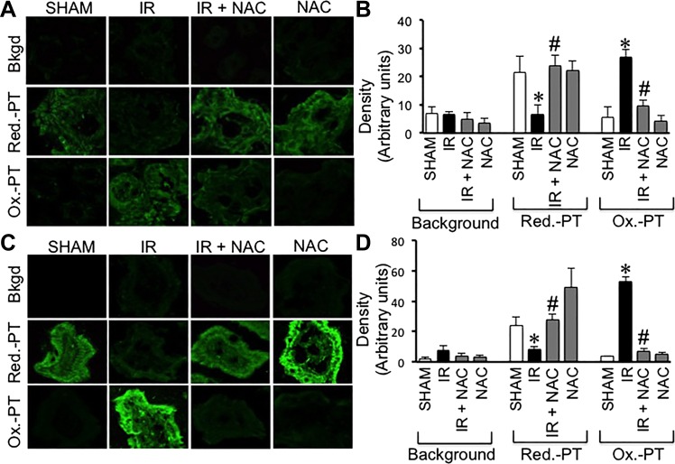
The goals of this study were to evaluate the effects of ionizing radiation on apical junctions in colonic epithelium and mucosal barrier function in mice in vivo. Adult mice were subjected to total body irradiation (4 Gy) with or without N-acetyl-l-cysteine (NAC) feeding for 5 days before irradiation. At 2-24 h postirradiation, the integrity of colonic epithelial tight junctions (TJ), adherens junctions (AJ), and the actin cytoskeleton was assessed by immunofluorescence microscopy and immunoblot analysis of detergent-insoluble fractions for TJ and AJ proteins. The barrier function was evaluated by measuring vascular-to-luminal flux of fluorescein isothiocyanate (FITC)-inulin in vivo and luminal-to-mucosal flux in vitro. Oxidative stress was evaluated by measuring protein thiol oxidation. Confocal microscopy showed that radiation caused redistribution of occludin, zona occludens-1, claudin-3, E-cadherin, and β-catenin, as well as the actin cytoskeleton as early as 2 h postirradiation, and this effect was sustained for at least 24 h. Feeding NAC before irradiation blocked radiation-induced disruption of TJ, AJ, and the actin cytoskeleton. Radiation increased mucosal permeability to inulin in colon, which was blocked by NAC feeding. The level of reduced-protein thiols in colon was depleted by radiation with a concomitant increase in the level of oxidized-protein thiol. NAC feeding blocked the radiation-induced protein thiol oxidation. These data demonstrate that radiation rapidly disrupts TJ, AJ, and the actin cytoskeleton by an oxidative stress-dependent mechanism that can be prevented by NAC feeding.

Keywords: actin; adherens junction; barrier function; cytoskeleton; intestine; occludin; oxidative stress; protein thiol; zona occludens-1.


Figures

Fig. 1.
Fig. 1.
Temporal effect of γ-irradiation (γ-IR) on colonic mucosal morphology in mice. Mice were subjected to total body irradiation [TBI (IR)] or sham treatment (Sham). At 2 or 24 h postirradiation (post-IR), sections of formalin-fixed distal colon were stained with hematoxylin and eosin. Bright-field images were captured.
Fig. 2.
Fig. 2.
γ-IR induces rapid redistribution of tight junction (TJ) proteins in mouse intestine. Mice were subjected to TBI (IR) or sham treatment (Sham). At 2–24 h post-IR, cryosections of distal colon (A) and ileum (B) were stained for occludin (green in A and red in B) and zona occludens (ZO)-1 (red in A and green in B) by the immunofluorescence method, and nuclei were stained with Hoechst 33342 (blue). Fluorescence images were captured by confocal microscopy.
Fig. 3.
Fig. 3.
γ-IR induces rapid redistribution of claudin (Cldn)-3 and reorganization of the actin cytoskeleton. Mice were subjected to TBI (IR) or sham treatment (Sham). At 2–24 h post-IR, cryosections of distal colon (A) and ileum (B) were stained for Cldn-3 (red) and F-actin (green) by the immunofluorescence method, and the nuclei were stained using Hoechst 33342 (blue).
Fig. 4.
Fig. 4.
γ-IR induces rapid redistribution of adherens junction (AJ) proteins in mouse intestine. Mice were subjected to TBI (IR) or sham treatment (Sham). At 2–24 h post-IR, cryosections of distal colon (A) and ileum (B) were stained for E-cadherin (green) and β-catenin (red) by the immunofluorescence method, and the nuclei were stained with Hoechst 33342 (blue).
Fig. 5.
Fig. 5.
N-acetyl-l-cysteine (NAC) treatment on colonic mucosal morphology in mice. Mice were fed a liquid diet with or without 20 mM NAC for 5 days before a 4 Gy dose of TBI (IR) or sham treatment (Sham). At 2 h post-IR, sections of formalin-fixed distal colon were stained with hematoxylin and eosin. Bright-field images were captured.
Fig. 6.
Fig. 6.
NAC feeding blocks γ-IR-induced redistribution of TJ proteins in mouse intestine. Mice were fed a liquid diet with or without 20 mM NAC for 5 days before a 4 Gy dose of TBI (IR) or sham treatment (Sham). At 2 h post-IR, cryosections of distal colon (A) and ileum (B) were stained for occludin (green) and ZO-1 (red) by the immunofluorescence method, and the nuclei were stained with Hoechst 33342 (blue).
Fig. 7.
Fig. 7.
NAC feeding blocks γ-IR-induced redistribution of Cldn-3 and reorganization of actin cytoskeleton. Mice were fed a liquid diet with or without 20 mM NAC for 5 days before TBI (IR) or sham treatment (Sham). At 2 h post-IR, cryosections of distal colon (A) and ileum (B) were stained for F-actin (green) and Cldn-3 (red) by the immunofluorescence method, and the nuclei were stained with Hoechst 33342 (blue).
Fig. 8.
Fig. 8.
NAC feeding blocks γ-IR-induced redistribution of AJ proteins in mouse intestine. Mice were fed a liquid diet with or without 20 mM NAC for 5 days before TBI (IR) or sham treatment (Sham). At 2 h post-IR, cryosections of distal colon (A) and ileum (B) were stained for E-cadherin (green) and β-catenin (red) by the immunofluorescence method, and the nuclei were stained with Hoechst 33342 (blue).
Fig. 9.
Fig. 9.
NAC feeding blocks γ-IR-induced depletion of AJ proteins in mouse intestine. Mice were fed a liquid diet with or without 20 mM NAC for 5 days before TBI (IR) or sham treatment (Sham). At 2 h post-IR, Triton-insoluble fractions were prepared from the distal colonic mucosa and immunoblotted for TJ and AJ proteins (A). Immunoblot bands for occludin (B), ZO-1 (C), Cldn-3 (D), E-cadherin (E), and β-catenin (F) were quantitated by densitometric analysis and normalized to band density of corresponding actin bands. Values are means ± SE (n = 3). *Significantly (P < 0.05) different from values for the corresponding control group. #Significantly (P < 0.05) different from values for the corresponding IR group.
Fig. 10.
Fig. 10.
NAC feeding blocks γ-IR-induced mucosal barrier dysfunction in mouse intestine. Adult mice were fed liquid diet with or without 20 mM NAC for 5 days before TBI (IR) or sham treatment (Sham). At 2 h post-IR, intestinal mucosal barrier function was evaluated by measuring vascular-to-luminal flux of fluorescein isothiocyanate (FITC)-inulin in vivo (A) and in vitro (B) as described in materials and methods. A: in vivo flux values are means ± SE (n = 6). *Significantly (P < 0.05) different from corresponding value for Sham-treated mice. #Significantly different from corresponding value for the IR group. B: at 3 h post-IR inulin absorption from the lumen of colonic loops was measured. Values for absorbed fluorescence are means ± SE (n = 5). *Significantly (P < 0.05) different from corresponding value for Sham-treated mice. #Significantly different from corresponding value for the IR group.
Fig. 11.
Fig. 11.
NAC feeding blocks γ-IR-induced protein thiol oxidation in mouse intestine. Adult mice were fed liquid diet with or without 20 mM NAC for 5 days before TBI (IR) or sham treatment (Sham). At 2 h post-IR, the levels of reduced and oxidized protein thiols in distal colon (A and B) and ileum (C and D) were measured as described in materials and methods. Fluorescence images were collected by confocal microscopy (A and C). Fluorescence density was evaluated by using Image J software (B and D). Values are means ± SE (n = 5). *Significantly (P < 0.05) different from corresponding value for Sham-treated mice. #Significantly different from corresponding value for the IR group.

References

    1. Anderson JM, Van Itallie CM. Physiology and function of the tight junction. Cold Spring Harb Perspect Biol 1: a002584, 2009. - PMC - PubMed
    1. Aruoma OI, Halliwell B, Hoey BM, Butler J. The antioxidant action of N-acetylcysteine: its reaction with hydrogen peroxide, hydroxyl radical, superoxide, and hypochlorous acid. Free Radic Biol Med 6: 593–597, 1989. - PubMed
    1. Baier JE, Neumann HA, Moeller T, Kissler M, Borchardt D, Ricken D. Radiation protection through cytokine release by N-acetylcysteine. Strahlenther Onkol 172: 91–98, 1996. - PubMed
    1. Baum B, Georgiou M. Dynamics of adherens junctions in epithelial establishment, maintenance, and remodeling. J Cell Biol 192: 907–917, 2011. - PMC - PubMed
    1. Cameron S, Schwartz A, Sultan S, Schaefer IM, Hermann R, Rave-Frank M, Hess CF, Christiansen H, Ramadori G. Radiation-induced damage in different segments of the rat intestine after external beam irradiation of the liver. Exp Mol Pathol 92: 243–258, 2012. - PubMed

Publication types

MeSH terms