

Characterizing optical properties and spatial heterogeneity of human ovarian tissue using spatial frequency domain imaging
- PMID: 26822943
- PMCID: PMC4728740
- DOI: 10.1117/1.JBO.21.10.101402
Characterizing optical properties and spatial heterogeneity of human ovarian tissue using spatial frequency domain imaging
Abstract
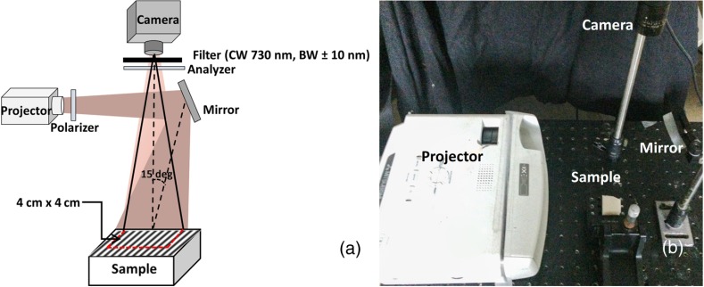
A spatial frequency domain imaging (SFDI) system was developed for characterizing ex vivo human ovarian tissue using wide-field absorption and scattering properties and their spatial heterogeneities. Based on the observed differences between absorption and scattering images of different ovarian tissue groups, six parameters were quantitatively extracted. These are the mean absorption and scattering, spatial heterogeneities of both absorption and scattering maps measured by a standard deviation, and a fitting error of a Gaussian model fitted to normalized mean Radon transform of the absorption and scattering maps. A logistic regression model was used for classification of malignant and normal ovarian tissues. A sensitivity of 95%, specificity of 100%, and area under the curve of 0.98 were obtained using six parameters extracted from the SFDI images. The preliminary results demonstrate the diagnostic potential of the SFDI method for quantitative characterization of wide-field optical properties and the spatial distribution heterogeneity of human ovarian tissue. SFDI could be an extremely robust and valuable tool for evaluation of the ovary and detection of neoplastic changes of ovarian cancer.
Figures


Similar articles
-
Hyperspectral imaging in the spatial frequency domain with a supercontinuum source.J Biomed Opt. 2019 Jul;24(7):1-9. doi: 10.1117/1.JBO.24.7.071614. J Biomed Opt. 2019. PMID: 31271005 Free PMC article.
-
Optical scattering coefficient estimated by optical coherence tomography correlates with collagen content in ovarian tissue.J Biomed Opt. 2011 Sep;16(9):090504. doi: 10.1117/1.3625247. J Biomed Opt. 2011. PMID: 21950907 Free PMC article.
-
Multi-frequency spatial frequency domain imaging: a depth-resolved optical scattering model to isolate scattering contrast in thin layers of skin.J Biomed Opt. 2024 Apr;29(4):046003. doi: 10.1117/1.JBO.29.4.046003. Epub 2024 Apr 22. J Biomed Opt. 2024. PMID: 38650893 Free PMC article.
-
An overview of optical coherence tomography for ovarian tissue imaging and characterization.Wiley Interdiscip Rev Nanomed Nanobiotechnol. 2015 Jan-Feb;7(1):1-16. doi: 10.1002/wnan.1306. Epub 2014 Oct 20. Wiley Interdiscip Rev Nanomed Nanobiotechnol. 2015. PMID: 25329515 Free PMC article. Review.
-
Non-invasive in vivo characterization of breast tumors using photon migration spectroscopy.Neoplasia. 2000 Jan-Apr;2(1-2):26-40. doi: 10.1038/sj.neo.7900082. Neoplasia. 2000. PMID: 10933066 Free PMC article. Review.
Cited by
-
A Comprehensive Study of Reactive Oxygen Species Explicit Dosimetry for Pleural Photodynamic Therapy.Antioxidants (Basel). 2024 Nov 22;13(12):1436. doi: 10.3390/antiox13121436. Antioxidants (Basel). 2024. PMID: 39765767 Free PMC article.
-
Ultrasound-enhanced Unet model for quantitative photoacoustic tomography of ovarian lesions.Photoacoustics. 2022 Oct 25;28:100420. doi: 10.1016/j.pacs.2022.100420. eCollection 2022 Dec. Photoacoustics. 2022. PMID: 36325304 Free PMC article.
-
Hyperspectral imaging in the spatial frequency domain with a supercontinuum source.J Biomed Opt. 2019 Jul;24(7):1-9. doi: 10.1117/1.JBO.24.7.071614. J Biomed Opt. 2019. PMID: 31271005 Free PMC article.
-
Reference-free determination of tissue absorption coefficient by modulation transfer function characterization in spatial frequency domain.Biomed Eng Online. 2017 Aug 8;16(1):100. doi: 10.1186/s12938-017-0394-z. Biomed Eng Online. 2017. PMID: 28789661 Free PMC article.
-
Virus-Mimicking Nanoparticles for Targeted Near Infrared Fluorescence Imaging of Intraperitoneal Ovarian Tumors in Mice.Ann Biomed Eng. 2021 Feb;49(2):548-559. doi: 10.1007/s10439-020-02589-8. Epub 2020 Aug 6. Ann Biomed Eng. 2021. PMID: 32761557
References
Publication types
MeSH terms
Grants and funding
LinkOut - more resources
Full Text Sources
Other Literature Sources
Medical
