. 2016 Jan 29:16:36.
doi: 10.1186/s12906-016-1015-2.

Assessment of cytotoxicity and genotoxicity of stem bark extracts from Canarium odontophyllum Miq. (dabai) against HCT 116 human colorectal cancer cell line

Affiliations

Assessment of cytotoxicity and genotoxicity of stem bark extracts from Canarium odontophyllum Miq. (dabai) against HCT 116 human colorectal cancer cell line

Dayang Fredalina Basri et al. BMC Complement Altern Med. .

Abstract

Background: Canarium odontophyllum Miq. is a plant species widely known as 'dabai' and can be vastly found in Sarawak. The aim of this study was to assess the cytotoxic and genotoxic effect of extracts from stem bark of C. odontophyllum against HCT 116 human colorectal cancer cell line.

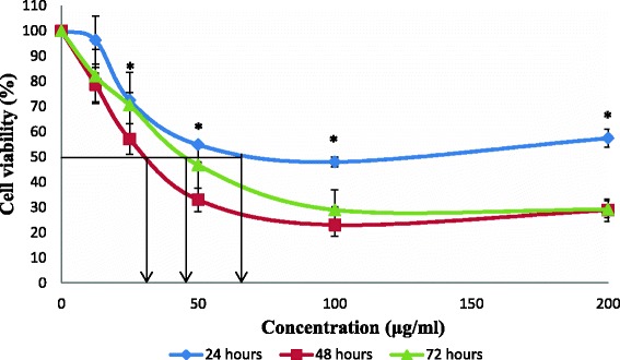
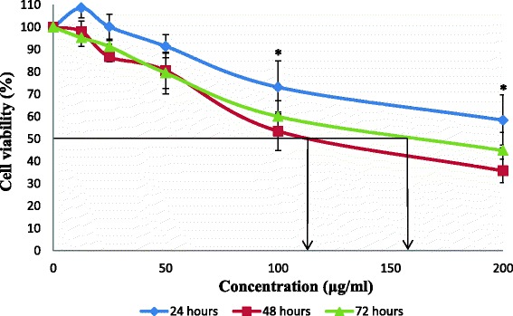
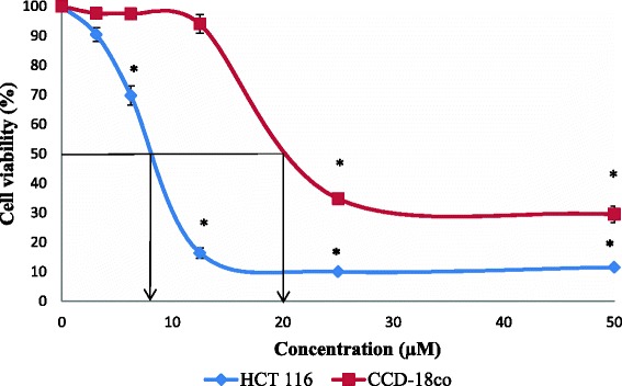
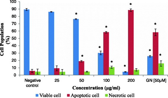
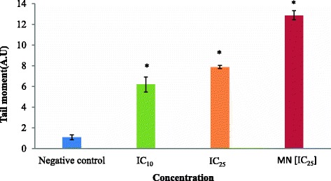
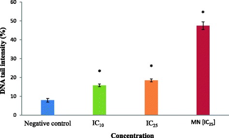
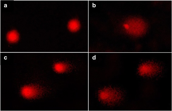
Method: The IC50 values of the aqueous, methanol, and acetone extracts against HCT 116 cells as well as the acetone extract against human colon fibroblast cell line CCD-18co were determined using the MTT assay. The concentration of the extracts ranged from 12.5 to 200 μg/ml at treatment time of 24, 48 and 72 h. Annexin V-FITC/PI labelling assay was employed to determine mode of HCT 116 cell death induced by acetone extract at 48 h. The DNA damage induced by the extract in HCT 116 cells was detected using alkaline comet assay at 30 min of IC10 and IC25 treatment.

Results: Acetone extract exhibited the highest cytotoxic effect against HCT 116 cells compared to methanol and aqueous extract at 24, 48 and 72 h. Despite no cytotoxic effect by acetone extract against CCD-18co cells at 24 and 48 h, however at 72 h, CCD-18co cells proliferated. Apoptosis assessment using Annexin V-FITC/PI labelling assay revealed that the primary cell death was via apoptosis after 48 h treatment. Low doses of acetone extract from stem bark of C. odontophyllum showed significant DNA damage in HCT 116 cells with tail moment of 6.187 ± 0.718 A.U and 7.877 ± 0.142 A.U, respectively.

Conclusions: Acetone extract from stem bark of C. odontophyllum has high potential in the development of anticancer agent against HCT 116 cells with no cytotoxic effect against human colon fibroblast cells.


Figures

Fig. 1
Fig. 1
Cell viability of HCT 116 cells after exposure of acetone extract from stem bark of C. odontophyllum. Cell survival is expressed as a percentage relative to the negative control (untreated). Acetone extract from stem bark of C. odontophyllum at concentration ranging from 0 – 200 μg/ml exhibited cytotoxic activity against HCT 116 cells at 24, 48 and 72 h of treatment exposure. Each point represents the mean of triplicates from 3 different experiments ± S.E.M
Fig. 2
Fig. 2
Cell viability of HCT 116 cells after exposure of methanol extract from stem bark of C. odontophyllum. Cell survival is expressed as a percentage relative to the negative control (untreated). Methanol extract from stem bark of C. odontophyllum at concentration ranging from 0 – 200 μg/ml exhibited cytotoxic activity against HCT 116 cells at 24, 48 and 72 h of treatment exposure. Each point represents the mean of triplicates from 3 different experiments ± S.E.M
Fig. 3
Fig. 3
Cell viability of HCT 116 cells after exposure of aqueous extract from stem bark of C. odontophyllum. Cell survival is expressed as a percentage relative to the negative control (untreated). Aqueous extract from stem bark of C. odontophyllum at concentration ranging from 0 – 200 μg/ml exhibited cytotoxic activity against HCT 116 cells at 24, 48 and 72 h of treatment exposure. Each point represents the mean of triplicates from 3 different experiments ± S.E.M
Fig. 4
Fig. 4
Cell viability of HCT 116 and CCD-18co cells after exposure of Menadione. Cell survival is expressed as a percentage relative to the negative control (untreated). Menadione at concentration ranging from 0 – 50 Μm exhibited cytotoxic activity against HCT 116 and CCD-18co cells at 24 h of treatment exposure. Each point represents the mean of triplicates from 3 different experiments ± S.E.M
Fig. 5
Fig. 5
Cell viability of CCD-18co cells after exposure of acetone extract from stem bark of C. odontophyllum. Cell survival is expressed as a percentage relative to the negative control (untreated). Acetone extract from stem bark of C. odontophyllum at concentration ranging from 0 – 200 μg/ml exhibited no cytotoxic activity against CCD-18co cells at 24, 48 and 72 h of treatment exposure. Each point represents the mean of triplicates from 3 different experiments ± S.E.M
Fig. 6
Fig. 6
Cytogram of HCT 116 cells induced with acetone extract. Cells were treated with acetone extract from stem bark of C. odontophyllum at concentration ranging from 25 – 200 μg/ml after 48 h of treatment
Fig. 7
Fig. 7
Percentage of cell population of HCT 116 after 48 h treatment. Cells were treated with of acetone extract from stem bark of C. odontophyllum at concentration ranging from 25 – 200 μg/ml for 48 h. Each data represents the mean of triplicates from 3 different experiments ± S.E.M
Fig. 8
Fig. 8
Tail moment of HCT 116 cells after 30 min of treatment. Cells were treated with acetone extract from stem bark of C. odontophyllum for 30 min. Each data represents the mean of triplicates from 3 different experiments ± S.E.M
Fig. 9
Fig. 9
DNA tail intensity of HCT 116 cells after 30 min of treatment. Cells were treated with acetone extract from stem bark of C. odontophyllum for 30 min. Each data represents the mean of triplicates from 3 different experiments ± S.E.M
Fig. 10
Fig. 10
Photo of a single HCT 116 cell stained with ethidium bromide under fluorescent microscope at 30 min. a Negative control: untreated cell (b) Menadione at IC25 (c) DNA migration of HCT 116 cell induced by acetone extract at IC10 (d) DNA migration of HCT 116 cell induced by acetone extract at IC25

References

    1. Underwood JCE, Cross SS. General and Systemic Pathology. Churchill Livingstone: Elsevier; 2009. p. 395.
    1. Cancer Genome Atlas Network Comprehensive molecular characterization of human colon and rectal cancer. Nature. 2012;487(7407):330–7. doi: 10.1038/nature11252. - DOI - PMC - PubMed
    1. Malaysian Gastrointestinal Registry Report 2009. 2009. http://www.crc.gov.my/wpcontent/uploads/documents/report/20100301_nerReport. Accessed 5 September 2014.
    1. Creţu E, Trifan A, Vasincu A, Miron A. Plant-derived anticancer agents-curcumin in cancer prevention and treatment. Rev Med Chir Soc Med Nat lasi. 2012;116(4):1223–9. - PubMed
    1. Ahmed M, Khan MI, Khan MR, Muhammad N, Khan AU. Role of medicinal plants in oxidative stress and cancer. Open Access Sci Rep. 2013;2(2):641.

Publication types