. 2016 Mar 15;27(6):1040-50.
doi: 10.1091/mbc.E15-07-0527. Epub 2016 Jan 28.

Regulatory roles of conserved phosphorylation sites in the activation T-loop of the MAP kinase ERK1

Affiliations

Regulatory roles of conserved phosphorylation sites in the activation T-loop of the MAP kinase ERK1

Shenshen Lai et al. Mol Biol Cell. .

Abstract

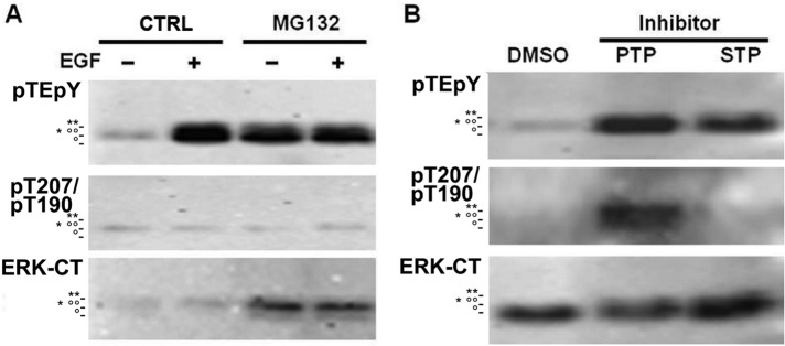
The catalytic domains of most eukaryotic protein kinases are highly conserved in their primary structures. Their phosphorylation within the well-known activation T-loop, a variable region between protein kinase catalytic subdomains VII and VIII, is a common mechanism for stimulation of their phosphotransferase activities. Extracellular signal-regulated kinase 1 (ERK1), a member of the extensively studied mitogen-activated protein kinase (MAPK) family, serves as a paradigm for regulation of protein kinases in signaling modules. In addition to the well-documented T202 and Y204 stimulatory phosphorylation sites in the activation T-loop of ERK1 and its closest relative, ERK2, three additional flanking phosphosites have been confirmed (T198, T207, and Y210 from ERK1) by high-throughput mass spectrometry. In vitro kinase assays revealed the functional importance of T207 and Y210, but not T198, in negatively regulating ERK1 catalytic activity. The Y210 site could be important for proper conformational arrangement of the active site, and a Y210F mutant could not be recognized by MEK1 for phosphorylation of T202 and Y204 in vitro. Autophosphorylation of T207 reduces the catalytic activity and stability of activated ERK1. We propose that after the activation of ERK1 by MEK1, subsequent slower phosphorylation of the flanking sites results in inhibition of the kinase. Because the T207 and Y210 phosphosites of ERK1 are highly conserved within the eukaryotic protein kinase family, hyperphosphorylation within the kinase activation T-loop may serve as a general mechanism for protein kinase down-regulation after initial activation by their upstream kinases.


Figures

FIGURE 1:
FIGURE 1:
Phosphorylation sites in human protein kinase catalytic domains. (A) Distribution of experimentally confirmed phosphosites in human protein kinase domains. Phosphosites identified in human protein kinase catalytic domains were mapped on the alignment provided in Supplemental Table S1. The total number of phosphosites was 1950, with 304 activatory sites (dark purple) mostly clustering at activation loop between aligned amino acid residues 139 and 159, and inhibitory sites (red). (B) Distribution of phosphotyrosine (orange), phosphothreonine (green), and phosphoserine (blue) residues in the kinase activation T-loop. The locations of the phosphorylation sites in ERK1 are shown in purple. These include the highly conserved threonine phosphorylation site at the aligned position 155 and tyrosine phosphorylation site at the aligned position 158. The T198 phosphosite of ERK1 is located with the Insert/Gap 8 region at aligned position 148.
FIGURE 2:
FIGURE 2:
Phosphorylation of ERK1 T207 and Y210 in vitro. (A) ERK1-WT and KD phosphorylation by MEK1-ΔN3EE. The reactions were carried out at 30°C for 15 min. (B) Time-course experiment of ERK1-WT phosphorylation. At each time point, an aliquot of the incubation mix was taken and mixed with SDS–PAGE sample buffer to terminate the reaction. The samples were subsequently probed with phosphosite-specific antibodies (ERK1 pT207, PYKSD8 for pY210 and dual phospho-ERK1/2 pTEpY for pT202 and pY204) or the pan-expression ERK1/2-CT (ERK-CT) antibody. Western blots from the region of the migration of ERK1. Similar results were obtained in at least three independent experiments.
FIGURE 3:
FIGURE 3:
Phosphorylation and activity of ERK1 and its mutants. Six ERK1 mutants were created to characterize the functional roles of the three flanking phosphosites near the TEY motif (A). Purified recombinant ERK1 and its mutants were incubated with MEK1-ΔN3EE (orange) or kinase dilution buffer (blue) in presence of 50 μM ATP at 30°C for 15 min. An aliquot of each reaction mix was mixed with 2.5 μg of MBP and incubated for another 2 min. Samples were mixed with SDS–PAGE sample buffer and analyzed by Western blotting using the dual phospho-ERK1/2 pTEpY antibody (B) and phospho-MBP antibody (C). The results are averaged from three to five separate experiments with the SDs indicated by bars. **p < 0.005.
FIGURE 4:
FIGURE 4:
Phosphorylation and activity of ERK1-WT, T207A, and T207E in HEK293 cells. (A) Phosphorylation of TEY motif of ERK1 under serum stimulation. HEK293 cells stably expressing Flag-ERK1 were starved overnight before stimulation with 10% FBS for 10 min. (B) Activity of immunoprecipitated Flag-ERK1. After serum stimulation, Flag-ERK1 was immunoprecipitated by Flag-tag antibody and incubated with 5 μg of MBP and 50 μM ATP at 30°C for 15 min. The samples were subsequently subjected to SDS–PAGE and Western blotting with phosphosite-specific antibodies for the dual phospho-ERK1/2 phosphosite pTEpY and myelin basic protein (pMBP) and the Flag tag (Flag). (C) Each image is representative of three independent experiments, and the averages of the phosphorylation of the pTEpY site and MBP from the three separate experiments are shown with the SDs indicated by bars. **p < 0.005.
FIGURE 5:
FIGURE 5:
Phosphorylation of ERK T207/T190 is regulated by protein phosphatases in A431 cells. (A) A431 cells were treated with 10 μM proteasome inhibitor MG132 for 4 h, followed by stimulation with 100 ng/ml EGF for 5 min. (B) A431 cells were treated with 0.025% dimethyl sulfoxide (CTRL), PTP inhibitors (25 μM PAO and 50 μM Na3VO4), or STP inhibitor (30 mM NaF) for 30 min. The samples were subsequently subjected to SDS–PAGE and Western blotting with antibodies that were dual phospho-ERK1/2 pTEpY phosphosite-specific and ERK1 pT207 phosphosite-specific (which cross-reacts with ERK2 pT190) or the pan-expression ERK1/2-CT (ERK-CT) antibody. The migration positions of phospho-ERK1 (**), phospho-ERK2 (°°), ERK1 (*), and ERK2 (°) on the SDS–PAGE gel are indicated.
FIGURE 6:
FIGURE 6:
Site-directed mutagenesis of phosphorylation sites in the activation T-loop of protein-serine/threonine kinases. Publications were identified in which site-directed mutagenesis had been performed on protein kinases that featured a phosphorylated threonine residue at aligned position 155 and/or a phosphorylated tyrosine residue at aligned position 158 in their catalytic domains. The effects of mutation of these and flanking phosphosites are shown as gain of function (GoF), loss of function (LoF), or without known effect (NoE) on the phosphotransferase activities of the tested protein kinases. Activatory phosphosites are highlighted in green, and suspected inhibitory phosphosites are highlighted in pink. A more complete list of mutated phosphorylation sites in diverse human proteins is provided in Supplemental Table S3B.
FIGURE 7:
FIGURE 7:
Interactions with T207 and Y210 residues in the 3D structure of human ERK1. The x-ray crystallographic structure of T204 phosphorylated human ERK1 (PDB Id 2ZOQ) was originally deduced by Kinoshita et al. (2008) and is rendered with JMol on the RCSB PDB website. The backbone atoms of ERK1 appear with white bonds, and most of the atoms in the tyrosine, tryptophan, and proline side-chain residues are colored orange, green, and yellow, respectively. Most oxygen, nitrogen, and carbon atoms in the other amino acid residue side chains appear as red, blue, and gray, respectively. Distances of atoms in the side chains of T207 and Y210 that were within 5 Å of the atoms of other amino acid side chains are indicated with orange dashed lines. In particular, T207 appears to interact with K168, D166, and Y210, whereas Y210 also interacts with P169, E237, and W209. Supplemental Table S4, A and B, respectively, provides listings of the actual distances between the phosphoacceptor residues in ERK1 and ERK2 with neighboring amino acid residues.

References

    1. Ahn NG, Seger R, Bratlien RL, Diltz CD, Tonks NK, Krebs EG. Multiple components in an epidermal growth factor-stimulated protein kinase cascade. In vitro activation of a myelin basic protein/microtubule-associated protein 2 kinase. J Biol Chem. 1991;266:4220–4227. - PubMed
    1. Alessi DR, Andjelkovic M, Caudwell B, Cron P, Morrice N, Cohen P, Hemmings BA. Mechanism of activation of protein kinase B by insulin and IGF-1. EMBO J. 1996;15:6541–6551. - PMC - PubMed
    1. Anderson NG, Maller JL, Tonks NK, Sturgill TW. Requirement for integration of signals from two distinct phosphorylation pathways for activation of MAP kinase. Nature. 1990;343:651–653. - PubMed
    1. Arnold R, Patzak IM, Neuhaus B, Vancauwenbergh S, Veillette A, Van Lint J, Kiefer F. Activation of hematopoietic progenitor kinase 1 involves relocation, autophosphorylation, and transphosphorylation by protein kinase D1. Mol Cell Biol. 2005;25:2364–2383. - PMC - PubMed
    1. Beullens M, Vancauwenbergh S, Morrice N, Derua R, Ceulemans H, Waelkens E, Bollen M. Substrate specificity and activity regulation of protein kinase MELK. J Biol Chem. 2005;280:40003–40011. - PubMed

Publication types

Substances

LinkOut - more resources