

Case Reports
doi: 10.4103/1008-682X.173440.
Rare primary seminal vesicle cystadenoma: computed tomography and magnetic resonance imaging findings
Affiliations
- PMID: 26823068
- PMCID: PMC5427799
- DOI: 10.4103/1008-682X.173440
Item in Clipboard
Case Reports
Rare primary seminal vesicle cystadenoma: computed tomography and magnetic resonance imaging findings
Asian J Androl.
2017 May-Jun.
No abstract available
Figures
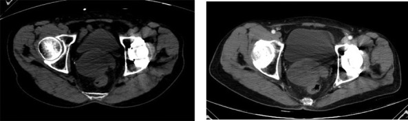
Pelvic CT scan shows a 5.5 cm × 6.0 cm × 5.3 cm sized oval, soft tissue density mass between the bladder and rectum. Unenhanced (a) and contrast-enhanced (b) CT scan show CT value of the tumor is about 10.5 HU and 24.1 HU, respectively CT: computed tomography.
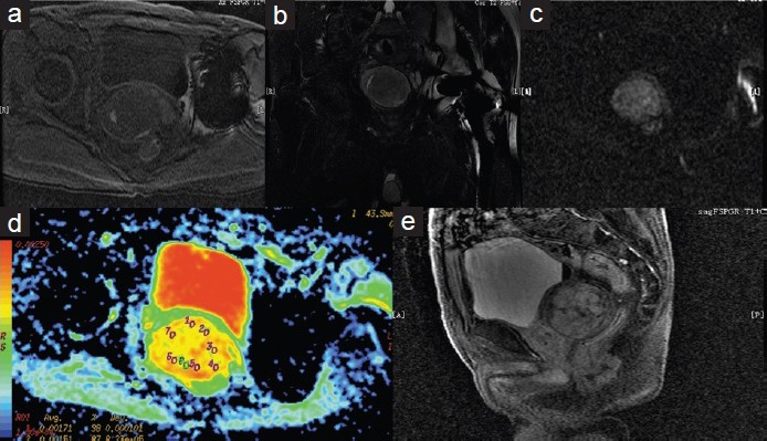
On cross section T1-weighted image (a), the tumor is mixed hypointense and hyperintense. On coronal T2-weighted (b) shows mixed hperintense signal tumor with hypointense capsule. On cross section, DW image (c) shows pale hyperintense signal. On cross section ADC image (d) shows the ADC is about 1.68 × 10⁻³ mm2 s−1. Contrast-enhanced MRI scan (e) shows prolonged and delayed enhancement pattern with tumor capsule. DW: diffusion-weighted; ADC: apparent diffusion coefficient; MRI: magnetic resonance imaging.
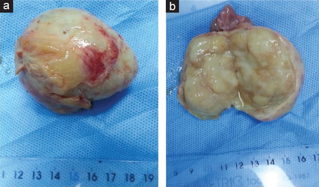
Grossly, the mass consisted of a well-circumscribed, oval contour. Its external surface is smooth, glistening (a). The cut surface showed multilocular cysts (b).
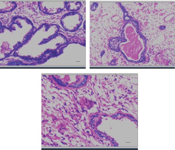
Microscopy: The tumor contains innumerable glands and cysts of varying sizes and shapes filled with pale eosinophilic intraluminal secretions and lined by one to two layers of cuboidal or low columnar cells. Scale bar = 100 μm (a and b). The stromal cells are spindle-shaped and arranged in fascicles between the glands. Scale bar = 200 μm (c).
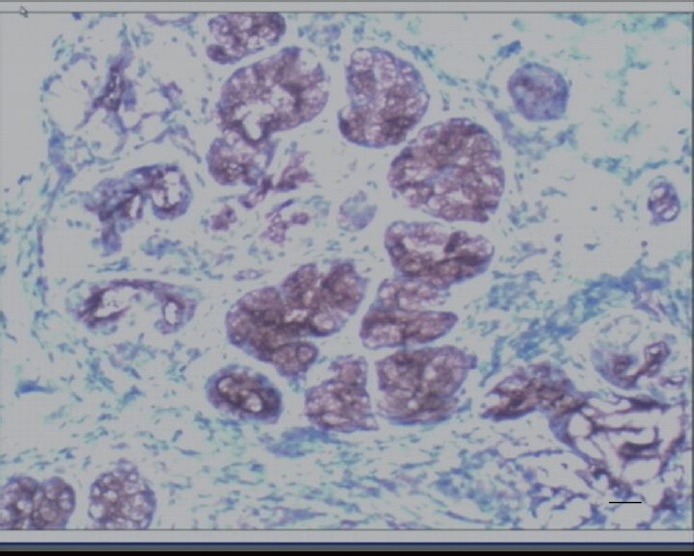
The glandular cells showed positivity for CA125. Scale bar = 100 μm.
Similar articles
-
Laparoscopic vesiculectomy for large seminal vesicle cystadenoma.Andrologia. 2019 Apr;51(3):e13209. doi: 10.1111/and.13209. Epub 2018 Nov 29. Andrologia. 2019. PMID: 30488974
-
Cystadenoma of the seminal vesicle: US and CT findings.Abdom Imaging. 1993;18(3):298-300. doi: 10.1007/BF00198130. Abdom Imaging. 1993. PMID: 8508100
-
Cystic schwannoma of a seminal vesicle.J Androl. 2012 Sep-Oct;33(5):798-800. doi: 10.2164/jandrol.111.015917. Epub 2012 Feb 23. J Androl. 2012. PMID: 22362077
-
Laparoscopic resection of a large mixed epithelial-stromal tumour of the seminal vesicle: a rare entity and review of the current literature.BMJ Case Rep. 2021 Feb 1;14(2):e238526. doi: 10.1136/bcr-2020-238526. BMJ Case Rep. 2021. PMID: 33526529 Free PMC article. Review.
-
[Cystadenoma of the seminal vesicles. A case report].Radiol Med. 1996 Mar;91(3):322-4. Radiol Med. 1996. PMID: 8628954 Review. Italian. No abstract available.
Cited by
-
Multiple schwannoma of the seminal vesicle: A case report.Medicine (Baltimore). 2020 Aug 14;99(33):e21603. doi: 10.1097/MD.0000000000021603. Medicine (Baltimore). 2020. PMID: 32872015 Free PMC article.
References
-
- Gil AO, Yamakami LY, Genzini T. Cystadenoma of the seminal vesicle. Int Braz J Urol. 2003;29:434–6. - PubMed
-
- Lee CB, Choi HJ, Cho DH, Ha US. Cystadenoma of the seminal vesicle. Int J urol. 2006;13:1138–40. - PubMed
-
- Lorber G, Pizov G, Gofrit ON, Pode D. Seminal vesicle cystadenoma: a rare clinical perspective. Eur Urol. 2011;60:388–91. - PubMed
-
- Lagalla R, Zappasodi F, Lo Casto A, Zenico T. Cystadenoma of the seminal vesicle: US and CT findings. Abdom Imaging. 1993;18:298–300. - PubMed
-
- Arora A, Sharma S, Seth A. Unusual retrovesical cytic mass in a male patient. Urology. 2013;81:e23–4. - PubMed
Publication types
MeSH terms
LinkOut - more resources
Full Text Sources
Other Literature Sources
