

Muscarinic receptor subtypes differentially control synaptic input and excitability of cerebellum-projecting medial vestibular nucleus neurons
- PMID: 26823384
- PMCID: PMC4828279
- DOI: 10.1111/jnc.13554
Muscarinic receptor subtypes differentially control synaptic input and excitability of cerebellum-projecting medial vestibular nucleus neurons
Abstract
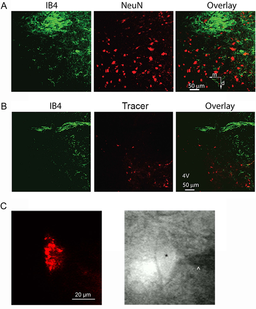
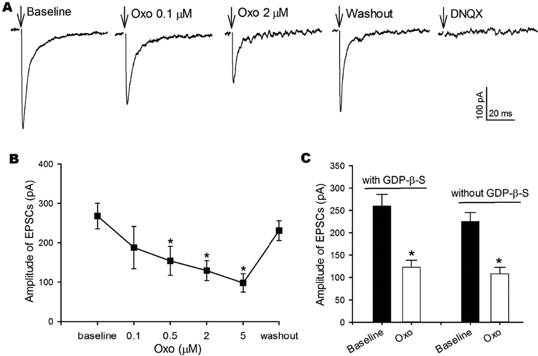
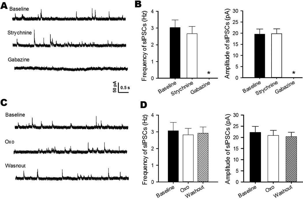
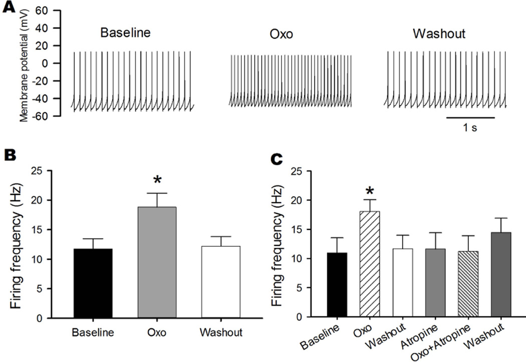
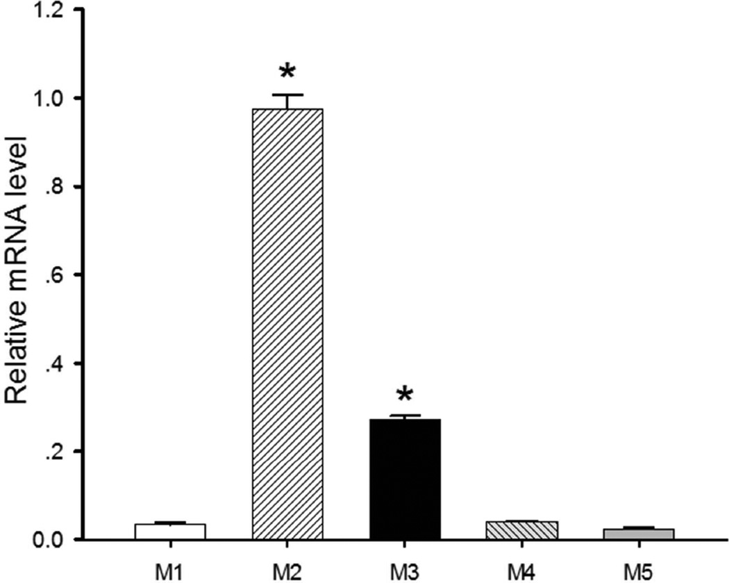
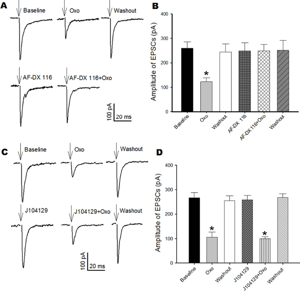
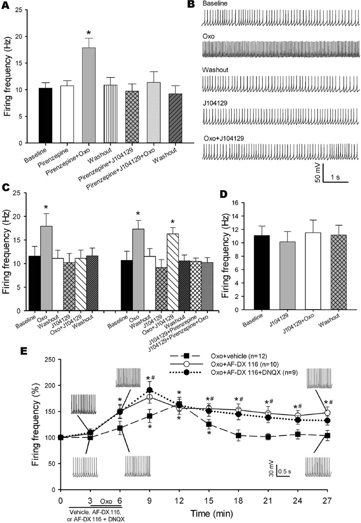
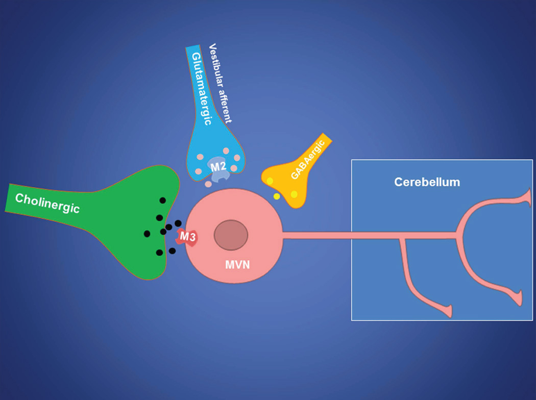
Neurons in the vestibular nuclei have a vital function in balance maintenance, gaze stabilization, and posture. Although muscarinic acetylcholine receptors (mAChRs) are expressed and involved in regulating vestibular function, it remains unclear how individual mAChR subtypes regulate vestibular neuronal activity. In this study, we determined which specific subtypes of mAChRs control synaptic input and excitability of medial vestibular nucleus (MVN) neurons that project to the cerebellum. Cerebellum-projecting MVN neurons were labeled by a fluorescent retrograde tracer and then identified in rat brainstem slices. Quantitative PCR analysis suggested that M2 and M3 were the possible major mAChR subtypes expressed in the MVN. The mAChR agonist oxotremorine-M significantly reduced the amplitude of glutamatergic excitatory post-synaptic currents evoked by stimulation of vestibular primary afferents, and this effect was abolished by the M2-preferring antagonist AF-DX 116. However, oxotremorine-M had no effect on GABA-mediated spontaneous inhibitory post-synaptic currents of labeled MVN neurons. Furthermore, oxotremorine-M significantly increased the firing activity of labeled MVN neurons, and this effect was blocked by the M3-preferring antagonist J104129 in most neurons tested. In addition, AF-DX 116 reduced the onset latency and prolonged the excitatory effect of oxotremorine-M on the firing activity of labeled MVN neurons. Our findings suggest that M3 is the predominant post-synaptic mAChR involved in muscarinic excitation of cerebellum-projecting MVN neurons. Pre-synaptic M2 mAChR regulates excitatory glutamatergic input from vestibular primary afferents, which in turn influences the excitability of cerebellum-projecting MVN neurons. This new information has important therapeutic implications for treating vestibular disorders with mAChR subtype-selective agents. Medial vestibular nucleus (MVN) neurons projecting to the cerebellum are involved in balance control. We found that activation of pre-synaptic M2 muscarinic receptors inhibit glutamatergic input from vestibular primary afferents, whereas stimulation of post-synaptic M3 muscarinic receptors increases the firing activity of cerebellum-projecting MVN neurons. This new information advances our understanding of the cholinergic mechanism regulating the vestibular system.
Keywords: acetylcholine; brainstem; glutamate; muscarinic receptors; synaptic transmission; vestibular system.
© 2016 International Society for Neurochemistry.
Conflict of interest statement
The authors have no conflict of interest to declare.
Figures
Similar articles
-
Regulation of glutamate release from primary afferents and interneurons in the spinal cord by muscarinic receptor subtypes.J Neurophysiol. 2007 Jan;97(1):102-9. doi: 10.1152/jn.00586.2006. Epub 2006 Oct 18. J Neurophysiol. 2007. PMID: 17050831
-
Muscarinic Acetylcholine Receptors and M-Currents Underlie Efferent-Mediated Slow Excitation in Calyx-Bearing Vestibular Afferents.J Neurosci. 2017 Feb 15;37(7):1873-1887. doi: 10.1523/JNEUROSCI.2322-16.2017. Epub 2017 Jan 16. J Neurosci. 2017. PMID: 28093476 Free PMC article.
-
Dynamic regulation of glycinergic input to spinal dorsal horn neurones by muscarinic receptor subtypes in rats.J Physiol. 2006 Mar 1;571(Pt 2):403-13. doi: 10.1113/jphysiol.2005.102905. Epub 2006 Jan 12. J Physiol. 2006. PMID: 16410279 Free PMC article.
-
Primary neurotransmitters and regulatory substances onto vestibular nucleus neurons.Biol Sci Space. 2001 Dec;15(4):371-4. doi: 10.2187/bss.15.371. Biol Sci Space. 2001. PMID: 12101361 Review.
-
Cholinergic innervation and receptors in the cerebellum.Prog Brain Res. 1997;114:67-96. doi: 10.1016/s0079-6123(08)63359-2. Prog Brain Res. 1997. PMID: 9193139 Review.
Cited by
-
α2δ-1-Dependent NMDA Receptor Activity in the Hypothalamus Is an Effector of Genetic-Environment Interactions That Drive Persistent Hypertension.J Neurosci. 2021 Jul 28;41(30):6551-6563. doi: 10.1523/JNEUROSCI.0346-21.2021. Epub 2021 Jun 30. J Neurosci. 2021. PMID: 34193557 Free PMC article.
-
Vestibular Functioning in Children with Neurodevelopmental Disorders Using the Functional Head Impulse Test.Brain Sci. 2020 Nov 20;10(11):887. doi: 10.3390/brainsci10110887. Brain Sci. 2020. PMID: 33233781 Free PMC article.
-
A comparative study of vestibular projection connectivity and balance in healthy young adults and elderly subjects.BMC Neurol. 2024 Sep 6;24(1):324. doi: 10.1186/s12883-024-03819-5. BMC Neurol. 2024. PMID: 39243007 Free PMC article.
-
LRRC8A-dependent volume-regulated anion channels contribute to ischemia-induced brain injury and glutamatergic input to hippocampal neurons.Exp Neurol. 2020 Oct;332:113391. doi: 10.1016/j.expneurol.2020.113391. Epub 2020 Jun 27. Exp Neurol. 2020. PMID: 32598930 Free PMC article.
-
Bipolar disorder in the balance.Eur Arch Psychiatry Clin Neurosci. 2019 Oct;269(7):761-775. doi: 10.1007/s00406-018-0935-x. Epub 2018 Aug 6. Eur Arch Psychiatry Clin Neurosci. 2019. PMID: 30083956
References
-
- Alvarez JC, Diaz C, Suarez C, Fernandez JA, Gonzalez del Rey C, Navarro A, Tolivia J. Neuronal loss in human medial vestibular nucleus. Anat Rec. 1998;251:431–438. - PubMed
-
- Barmack NH. Central vestibular system: vestibular nuclei and posterior cerebellum. Brain Res Bull. 2003;60:511–541. - PubMed
-
- Barmack NH, Baughman RW, Errico P, Shojaku H. Vestibular primary afferent projection to the cerebellum of the rabbit. J Comp Neurol. 1993;327:521–534. - PubMed
-
- Bevan P. [3H]oxotremorine-M binding to membranes prepared from rat brain and heart: evidence for subtypes of muscarinic receptors. Eur J Pharmacol. 1984;101:101–110. - PubMed
Publication types
MeSH terms
Substances
Grants and funding
LinkOut - more resources
Full Text Sources
Other Literature Sources
