. 2016 Feb 2;7(5):6281-93.
doi: 10.18632/oncotarget.7057.

Ki-67 is required for maintenance of cancer stem cells but not cell proliferation

Affiliations

Ki-67 is required for maintenance of cancer stem cells but not cell proliferation

Justin Cidado et al. Oncotarget. .

Abstract

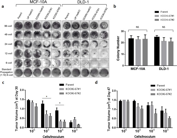
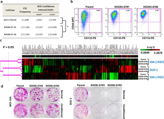
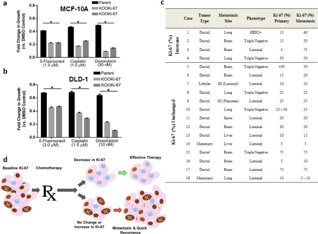
Ki-67 expression is correlated with cell proliferation and is a prognostic marker for various cancers; however, its function is unknown. Here we demonstrate that genetic disruption of Ki-67 in human epithelial breast and colon cancer cells depletes the cancer stem cell niche. Ki-67 null cells had a proliferative disadvantage compared to wildtype controls in colony formation assays and displayed increased sensitivity to various chemotherapies. Ki-67 null cancer cells showed decreased and delayed tumor formation in xenograft assays, which was associated with a reduction in cancer stem cell markers. Immunohistochemical analyses of human breast cancers revealed that Ki-67 expression is maintained at equivalent or greater levels in metastatic sites of disease compared to matched primary tumors, suggesting that maintenance of Ki-67 expression is associated with metastatic/clonogenic potential. These results elucidate Ki-67's role in maintaining the cancer stem cell niche, which has potential diagnostic and therapeutic implications for human malignancies.

Keywords: Ki-67; cancer stem cells; clonogenicity; proliferation; tumorigenicity.


Conflict of interest statement

CONFLICTS OF INTEREST

B.H.P. is a paid consultant for Novartis. B.H.P. is a paid member of the scientific advisory boards of Horizon Discovery, LTD and Loxo Oncology. Under separate licensing agreements between Horizon Discovery, LTD and The Johns Hopkins University, B.H.P. is entitled to a share of royalties received by the University on sales of products. The terms of this arrangement are being managed by the Johns Hopkins University in accordance with its conflict of interest policies. All other authors declare no potential conflicts.

Figures

Figure 1
Figure 1. Gene targeting of Ki-67
a. The targeting constructs consist of a neomycin resistance gene flanked by loxP sites and homology arms. Two promoterless adeno-associated virus (AAV) targeting vectors were synthesized, differing only in their 3′ homology arms termed “UP” and “DOWN” for relative 5′ and 3′ positions, respectively. Shown are constructs before Cre mediated excision. Red bars denote “STOP” sequences in all three reading frames. b. Targeting frequencies of individual UP and DOWN vectors in MCF-10A and DLD-1 cells. c. PCR was performed to confirm gene targeting of MKI67 alleles. The reverse primer was designed to anneal to a genomic sequence that is deleted (formula image
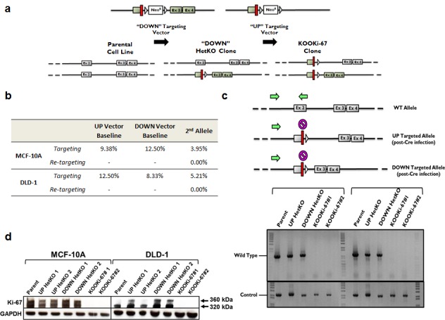
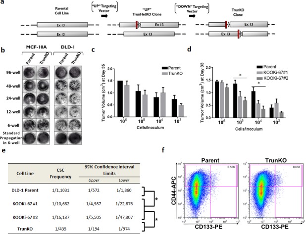
) with either vector. The absence of a PCR product for the KOOKi-67 clones indicates both alleles have been properly targeted. A separate control PCR across the first coding exon was performed to ensure the presence of gDNA in all samples. Green arrows denote primers used in PCR screens. d. Western blot for Ki-67 protein in parental, HetKO and KOOKi-67 cell lines using GAPDH as an internal loading control.
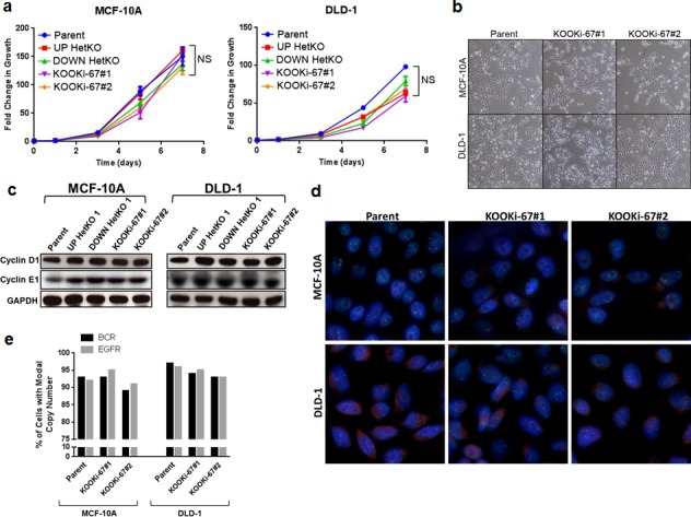
Figure 2
Figure 2. Loss of Ki-67 does not affect cell proliferation in bulk culture or alter morphology and does not induce chromosomal instability
a. MCF-10A and DLD-1 isogenic cell lines were seeded at 103 cells per well in 96-well plates to measure cell growth over a 7-day time course via CellTiter-Glo. NS = not significant. b. Representative phase contrast micrographs displaying normal cell morphology for MCF-10A and DLD-1 parental and KOOKi-67 clones (200x). c. Western blot for cyclin D1 and cyclin E1 in parental, HetKO and KOOKi-67 cell lines using GAPDH as an internal loading control. d. FISH was performed on parental and KOOKi-67 clones from MCF-10A and DLD-1 cells to assess for chromosomal instability. Cells were probed for EGFR (red) and BCR (green) loci, with representative experiments displayed. e. The modal copy number (N = 2 for both probes) was determined by counting 200 cells from each cell line, and chromosomal instability assessed as cells deviating from the modal copy number.
Figure 3
Figure 3. Ki-67 null cells have decreased clonogenic proliferation in vitro and in vivo
a. MCF-10A and DLD-1 isogenic cell lines were seeded at 103 cells per well in 96-well, 48-well, 24-well, 12-well, and 6-well plates under standard growth conditions, grown for 10 days, and stained with crystal violet. b. Parental and KOOKi-67 clones were also seeded at 75 cells per well in 6-well plates and grown for 10 days under standard conditions, at which time the number of colonies per well were counted. NS = not significant. c. DLD-1 parental and KOOKi-67 clones were injected subcutaneously into the flank of athymic female nude mice at the varying densities shown. Tumor volume was measured once per week until the tumor reached a maximal volume of 1.5 cm3. The average tumor volumes are plotted to display differences between parental and KOOKi-67 cells at each concentration used for injection at Day 30 and d. Day 47. * p < 0.05. All experiments are representative of at least three experiments using multiple replicates within each experiment.
Figure 4
Figure 4. Cancer stem cell frequency and markers are decreased in KOOKi-67 clones without global changes in protein expression
a. Stem cell frequency was calculated as described in the text using extreme limiting dilution analysis (ELDA); p = 0.000029. b. Flow cytometry was performed to assess the population of CD133+CD44+ cells in DLD-1 parental and KOOKi-67 cells. Representative results are shown from three independent experiments. c. MCF-10A and DLD-1 parental and KOOKi-67 clones were subjected to RPPA analysis. DLD-1 cells were grown in standard full growth conditions while MCF-10A cells were grown in both full growth (+EGF) and growth arrest (-EGF) condition. d. MCF-10A and DLD-1 parent cell lines were grown to 70 – 80% confluency, and conditioned media was filtered through a 0.2 μm filter. Parent and KOOKi-67 cell lines were seeded at 103 cells per well in 6-well plates and grown over 10 days in either normal growth media or a 1:1 mixture of conditioned:normal growth media.
Figure 5
Figure 5. The amino terminus of Ki-67 containing a FHA domain can regulate the cancer stem cell compartment
a. Schematic of targeting constructs used to create truncated knock outs (TrunKO). Two promoterless adeno-associated virus (AAV) targeting vectors were synthesized, designed to target distinct regions of the Ki-67 repeat domains and termed “UP” and “DOWN” for relative 5′ and 3′ positions, respectively. Shown are constructs after Cre mediated excision. Red bars denote “STOP” sequences in all three reading frames. b. MCF-10A and DLD-1 parental and TrunKO cell lines were seeded at 103 cells per well in 96-well, 48-well, 24-well, 12-well, and 6-well plates under standard growth conditions, grown for 10 days, and stained with crystal violet. c. DLD-1 parental, TrunKO and KOOKi-67 clones were injected subcutaneously into the flank of athymic female nude mice at the varying densities shown. Tumor volume was measured once per week until the tumor reached a maximal volume of 1.5 cm3. The average tumor volumes are plotted to display differences between parental, TrunKO and KOOKi-67 cells at each concentration used for injection. * p < 0.05. e. Stem cell frequency was calculated as described in the text using extreme limiting dilution analysis (ELDA); *p < 0.0001. f. Flow cytometry was performed to assess the population of CD133+CD44+ cells in DLD-1 parental and TrunKO cells.
Figure 6
Figure 6. Ki-67 expressing cells are relatively resistant to chemotherapy and persistent Ki-67 expression is present in metastatic tumors
a. Parental and KOOKi-67 cell lines from MCF-10A and b. DLD-1 cells were seeded and treated the following day with chemotherapies and vehicle control as described in Methods. The results are shown as fold change in growth relative to the vehicle control. * p<0.05. Results are representative from three independent experiments. c. Ki-67 staining of matched primary and metastatic breast cancers. HER2, human epidermal growth factor 2; GI, gastrointestinal tract. d. A proposed model of how changes in Ki-67 after chemotherapy may predict for effective versus ineffective response. Ki-67 positive cells (brown nuclei) mediate the stem cell (SC) niche. Effective chemotherapy dramatically reduces Ki-67 expressing cells, which in turn leads to reduced or eradicated cancer stem cells. Ineffective chemotherapy may reduce the bulk population of cells, but the percentage of Ki-67 expressing cells and subsequently stem cells remain unaffected, resulting in progression and metastases.

References

    1. Sasaki K, Murakami T, Kawasaki M, Takahashi M. The cell cycle associated change of the Ki-67 reactive nuclear antigen expression. J Cell Physiol. 1987;133:579–584. - PubMed
    1. Verheijen R, Kuijpers HJ, van Driel R, Beck JL, van Dierendonck JH, Brakenhoff GJ, Ramaekers FC. Ki-67 detects a nuclear matrix-associated proliferation-related antigen. II. Localization in mitotic cells and association with chromosomes. J Cell Sci. 1989;92:531–540. - PubMed
    1. Urruticoechea A, Smith IE, Dowsett M. Proliferation marker Ki-67 in early breast cancer. J Clin Oncol. 2005;23(28):7212–7220. - PubMed
    1. Paik S, Shak S, Tang G, Kim C, Baker J, Cronin M, Baehner FL, Walker MG, Watson D, Park T, Hiller W, Fisher ER, Wickerham DL, Bryant J, Wolmark N. A multigene assay to predict recurrence of tamoxifen-treated, node-negative breast cancer. N Engl J Med. 2004;351:2817–2826. - PubMed
    1. Van Bockstaele DR, Lan J, Snoeck HW, Korthout ML, De Bock RF, Peetermans ME. Aberrant Ki-67 expression in normal bone marrow revealed by multiparameter flow cytometric analysis. Cytometry. 1991:1250–63. - PubMed

Publication types

MeSH terms