

Spontaneous Reproductive Tract Lesions in Aged Captive Chimpanzees
- PMID: 26823448
- PMCID: PMC5607477
- DOI: 10.1177/0300985815620654
Spontaneous Reproductive Tract Lesions in Aged Captive Chimpanzees
Abstract
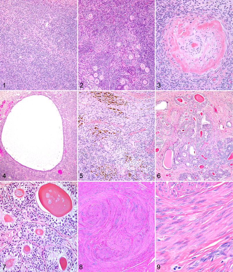
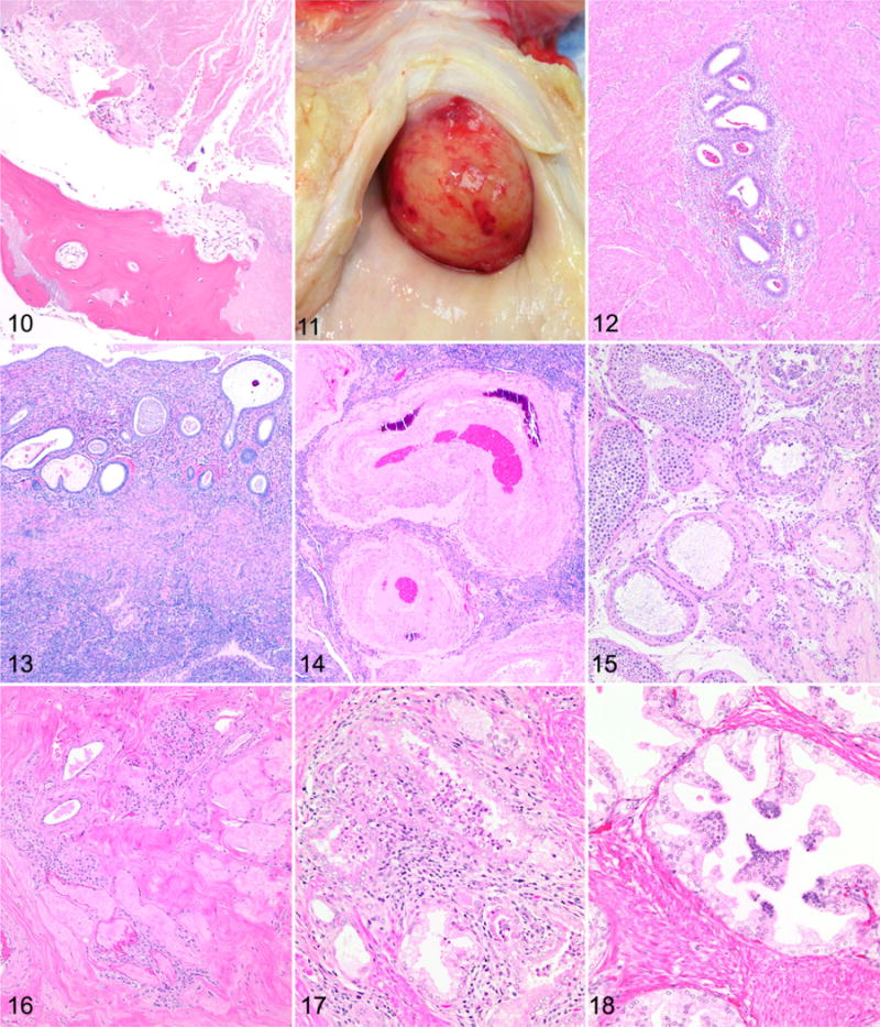
Chimpanzees (Pan troglodytes) have served as an important model for studies of reproductive diseases and aging-related disorders in humans. However, limited information is available about spontaneously occurring reproductive tract lesions in aging chimpanzees. In this article, the authors present histopathologic descriptions of lesions identified in the reproductive tract, including the mammary gland, of 33 female and 34 male aged chimpanzees from 3 captive populations. The most common findings in female chimpanzees were ovarian atrophy, uterine leiomyoma, adenomyosis, and endometrial atrophy. The most common findings in male chimpanzees were seminiferous tubule degeneration and lymphocytic infiltrates in the prostate gland. Other less common lesions included an ovarian granulosa cell tumor, cystic endometrial hyperplasia, an endometrial polyp, uterine artery hypertrophy and mineralization, atrophic vaginitis, mammary gland inflammation, prostatic epithelial hyperplasia, dilated seminal vesicles, a sperm granuloma, and lymphocytic infiltrates in the epididymis. The findings in this study closely mimic changes described in the reproductive tract of aged humans, with the exception of a lack of malignant changes observed in the mammary gland and prostate gland.
Keywords: biological aging; chimpanzee; female; genital organs; leiomyoma; male; mammary glands; pathology; uterine.
© The Author(s) 2016.
Conflict of interest statement
The author(s) declared no potential conflicts of interest with respect to the research, authorship, and/or publication of this article.
Figures
Similar articles
-
Natural pathology of the captive chimpanzee (Pan troglodytes): A 35-year review.J Med Primatol. 2017 Oct;46(5):271-290. doi: 10.1111/jmp.12277. Epub 2017 May 23. J Med Primatol. 2017. PMID: 28543059 Free PMC article. Review.
-
Comparative Pathology of Aging Great Apes: Bonobos, Chimpanzees, Gorillas, and Orangutans.Vet Pathol. 2016 Mar;53(2):250-76. doi: 10.1177/0300985815612154. Epub 2015 Dec 31. Vet Pathol. 2016. PMID: 26721908 Review.
-
Echocardiography parameters of clinically normal adult captive chimpanzees (Pan troglodytes).J Am Vet Med Assoc. 2014 Apr 15;244(8):956-60. doi: 10.2460/javma.244.8.956. J Am Vet Med Assoc. 2014. PMID: 24697773
-
A case of maxillary sarcoma in a chimpanzee (Pan troglodytes).J Med Primatol. 2014 Apr;43(2):111-4. doi: 10.1111/jmp.12086. Epub 2013 Dec 5. J Med Primatol. 2014. PMID: 24304143
-
Pathology of spontaneous air sacculitis in 37 baboons and seven chimpanzees and a brief review of the literature.J Med Primatol. 2012 Aug;41(4):266-77. doi: 10.1111/j.1600-0684.2012.00547.x. Epub 2012 Jul 6. J Med Primatol. 2012. PMID: 22765381 Free PMC article. Review.
Cited by
-
Hypermethylation of Klotho and Peroxisome Proliferator-Activated Receptor γ Concomitant with Overexpression of DNA Methyltransferase 1 in Adenomyosis.Reprod Sci. 2025 Mar;32(3):668-683. doi: 10.1007/s43032-024-01599-4. Epub 2024 May 30. Reprod Sci. 2025. PMID: 38816595
-
Urogenital Lesions in Nonhuman Primates at 2 National Primate Research Centers.Vet Pathol. 2021 Jan;58(1):147-160. doi: 10.1177/0300985820971752. Epub 2020 Nov 19. Vet Pathol. 2021. PMID: 33208023 Free PMC article.
-
Of Elephants and Other Mammals: A Comparative Review of Reproductive Tumors and Potential Impact on Conservation.Animals (Basel). 2022 Aug 8;12(15):2005. doi: 10.3390/ani12152005. Animals (Basel). 2022. PMID: 35953994 Free PMC article. Review.
-
Natural pathology of the captive chimpanzee (Pan troglodytes): A 35-year review.J Med Primatol. 2017 Oct;46(5):271-290. doi: 10.1111/jmp.12277. Epub 2017 May 23. J Med Primatol. 2017. PMID: 28543059 Free PMC article. Review.
-
Mpox infection of stromal cells and macrophages of macaque with endometriosis.Sci Rep. 2024 Sep 20;14(1):21947. doi: 10.1038/s41598-024-73012-8. Sci Rep. 2024. PMID: 39304769 Free PMC article.
References
-
- Archer DF, McIntyre-Seltman K, Wilborn WW, Jr, et al. Endometrial morphology in asymptomatic postmenopausal women. Am J Obstet Gynecol. 1991;165(2):317–320. - PubMed
MeSH terms
Grants and funding
LinkOut - more resources
Full Text Sources
Other Literature Sources
Medical
