. 2016 Apr 1;291(14):7703-15.
doi: 10.1074/jbc.M115.698282. Epub 2016 Jan 28.

Multiple Plasmodium falciparum Merozoite Surface Protein 1 Complexes Mediate Merozoite Binding to Human Erythrocytes

Affiliations

Multiple Plasmodium falciparum Merozoite Surface Protein 1 Complexes Mediate Merozoite Binding to Human Erythrocytes

Clara S Lin et al. J Biol Chem. .

Abstract

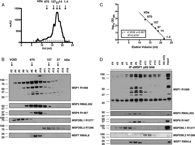
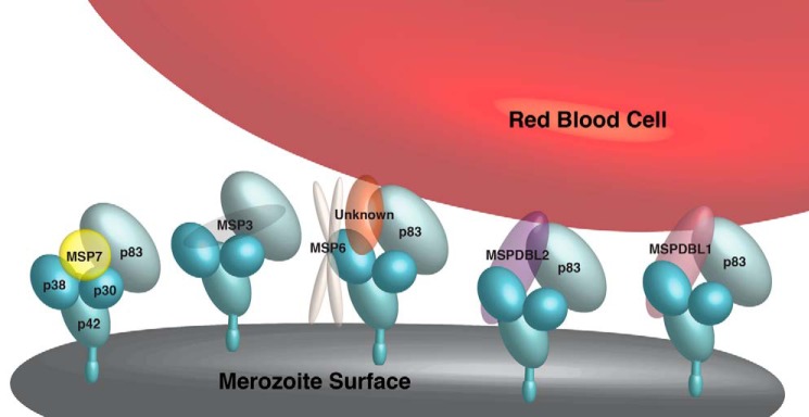
Successful invasion of human erythrocytes byPlasmodium falciparummerozoites is required for infection of the host and parasite survival. The early stages of invasion are mediated via merozoite surface proteins that interact with human erythrocytes. The nature of these interactions are currently not well understood, but it is known that merozoite surface protein 1 (MSP1) is critical for successful erythrocyte invasion. Here we show that the peripheral merozoite surface proteins MSP3, MSP6, MSPDBL1, MSPDBL2, and MSP7 bind directly to MSP1, but independently of each other, to form multiple forms of the MSP1 complex on the parasite surface. These complexes have overlapping functions that interact directly with human erythrocytes. We also show that targeting the p83 fragment of MSP1 using inhibitory antibodies inhibits all forms of MSP1 complexes and disrupts parasite growthin vitro.

Keywords: erythrocyte; infectious disease; invasion; malaria; protein complex.


Figures

FIGURE 1.
FIGURE 1.
Processed peripheral MSPs become part of an anchored complex late in schizogony. A, schematic showing the primary structure and processing sites of MSP1, MSP3, MSP6, MSPDBL1, MSPDBL2, and MSP7, with the final shed products indicated above the protein. Arrows indicate processing sites. Monoclonal antibodies with epitopes that target specific domains within the MSPs are shown (i.e. 9A6 (anti-MSP1 p83), 8D4 (anti-MSP1 p38), 5H7 (anti-MSP1 p30), 9A12 (anti-MSP3), 2C12 (anti-MSP6), 7H12 (anti-MSPDBL1), 4A7 (anti-MSPDBL2), and 8F1 (anti-MSP7)). B, immunofluorescence assays of trophozoites (Trophs, 28–32 h) in columns 1 (phase + 488 + DAPI) and 2 (488 + DAPI), schizonts (Schiz, 44–48 h) in columns 3 (phase + 488 + DAPI) and 4 (488 + DAPI), and PEMS (E64-treated) in columns 5 (phase + 488 + DAPI) and 6 (488 + DAPI) were probed with the monoclonal antibodies (green) 9A6 (anti-MSP1 p83), 9A12 (anti-MSP3), 2C12 (anti-MSP6), 7H12 (anti-MSPDBL1), 4A7 (anti-MSPDBL2), and 8F1 (anti-MSP7). EXP2 was used as an expression control probed with polyclonal rabbit anti-EXP2 antibodies (red). The nuclei of parasites were stained with DAPI (blue). C, parasites were harvested at three stages (trophozoites (28–32 h), schizonts (44–48 h), and PEMs (E64-treated)) together with the supernatant (SN) from invaded merozoites. Timing of processing and incorporation of MSPs onto a GPI-anchored MSP complex was observed through immunoblots of the three parasite stages, containing the supernatant fraction, which represents proteins that are in the parasitophorous vacuole, and the pellet fraction (represents proteins on the merozoite surface). The culture supernatant was released into the medium during merozoite invasion. Immunoblots for MSP1 p83, MSP1 p38, MSP1 p30, MSP3, MSP6, MSPDBL1, MSPDBL2, and MSP7 are shown, along with the monoclonal antibodies used, together with Hsp70 and AMA1, which is an expression control. Asterisks indicate nonspecific bands from red blood cell contaminants in the supernatant control, and arrows indicate the final processed products of the various MSPs.
FIGURE 2.
FIGURE 2.
MSP1 is the platform on the merozoite surface with which other MSPs interact. 3D7 parental parasite invasion supernatant was immunoprecipitated with monoclonal antibodies (9A6 (anti-MSP1), 2C12 (anti-MSP6), 7H12 (anti-MSPDBL1), or 4A7 (anti-MSPDBL2)). The first lane shows the invasion supernatant (Input) along with the eluates from immunoprecipitations (IP) of anti-MSP1, anti-MSP6, anti-MSPDBL1, or anti-MSPDBL2. The immunoblots were probed with the rabbit polyclonal antibodies R1566 (anti-MSP1), RMAL302 (anti-MSP3), R1467 (anti-MSP6), R1277 (anti-MSPDBL1), R1296 (anti-MSPDBL2), or RMAL8 (anti-MSP7). AMA1 was used as a negative control.
FIGURE 3.
FIGURE 3.
Recombinant MSP1, MSP3, MSP6, MSPDBL1, MSPDBL2, and MSP7 are recognized by human sera. A, final purified recombinant proteins for MSP1, MSP3, MSP6, MSPDBL1, MSPDBL2, and MSP7 are shown on a Coomassie-stained SDS-PAGE in the presence (+) and absence (−) of β-mercaptoethanol (βME). B, the recombinant proteins were probed on an immunoblot with pooled sera obtained from individuals infected by malaria in highly endemic regions in Papua New Guinea (aHyperimmune Sera).
FIGURE 4.
FIGURE 4.
The monoclonal antibodies against MSP1, MSP3, MSP6, MSPDBL1, MSPDBL2, and MSP7 used for immunoprecipitation assays are specific. Monoclonal antibodies against MSP1, MSP3, MSP6, MSPDBL1, MSPDBL2, MSP7, and MSP9 were screened for their specificity against native invasion complexes from parasites (left panels) and recombinant proteins (center panels) using immunoblots. The monoclonal antibodies were also screened with ELISAs (right panels) to show specificity to the antigen of interest. IS, invasion supernatant.
FIGURE 5.
FIGURE 5.
Recombinant MSPs interact with MSP1. A, recombinant proteins were incubated with recombinant MSP1 and immunoprecipitated (IP) with either anti-MSP1 monoclonal antibody or each of the monoclonal antibodies specific for MSP3, MSP6, MSPDBL1, MSPDBL2, or MSP7. The first and second lanes of each immunoblot represent the input of each recombinant protein and demonstrate that the antibodies are not cross-reactive. B, recombinant AMA1 immunoprecipitated with anti-AMA1 antibodies but did not co-immunoprecipitate with MSP1, MSP3, MSP6, MSPDBL1, MSPDBL2, or MSP7.
FIGURE 6.
FIGURE 6.
MSP1 complexes vary in size. A, gel filtration profile of concentrated invasion supernatant on Superose 6 10/300 GL with estimated molecular masses indicated by arrows. mAu, milli-absorbance units. B, immunoblots of Superose 6-fractionated invasion supernatant show that proteins MSP1, MSP3, MSP6, MSPDBL1, MSPDBL2, and MSP7 elute in different fractions at very high molecular masses. Molecular masses of standards are indicated by arrows. C, linear regression derived from molecular standards (thyroglobulin (670 kDa), bovine γ-globulin (158 kDa), chicken ovalbumin (44 kDa), equine myoglobulin (17 kDa), and vitamin B12 (1.4 kDa)) with elution volumes of 12.70, 15.92, 17.52, 18.83, and 21.59 ml, respectively. Approximate molecular mass was fitted to y = −0.3039 × +6.867, where R2 = 0.9797. D, Superose 6-fractionated samples 4–14 were immunoprecipitated (IP) with an anti-MSP1 monoclonal antibody (Ab), and eluates were probed with antibodies against MSP1, MSP3, MSP6, MSPDBL1, MSPDBL2, and MSP7.
FIGURE 7.
FIGURE 7.
MSP1 complexes have different roles in invasion. A, invasion supernatants harvested were allowed to bind to red blood cells, and the eluates were immunoblotted with specific antibodies against MSP1, MSP3, MSP6, MSPDBL1, MSPDBL2, and MSP7. The first lane represents the invasion supernatant input, the second lane is an RBC control that detects any nonspecific reactivity of antibodies to red blood cells, and the third lane is the eluate from the red blood cell binding assay. B, Superose 6-fractionated invasion supernatant (fractions 4–14) were incubated with host RBCs, and eluates were electrophoresed by SDS-PAGE and immunoblotted with either anti-MSP1, MSP3, MSP6, MSPDBL1, MSPDBL2, or MSP7 antibodies. PBS was used as an RBC control to control for nonspecific red blood cell proteins recognized by the antibodies. Proteins from invasion supernatant prefractionated served as molecular weight controls in the input lane. C, concentrated invasion supernatants were incubated with untreated RBCs (U) or RBCs treated with either chymotrypsin (C), trypsin (T), or neuraminidase (N). The complexes that contained MSP1, MSP6, MSPDBL1, and MSPDBL2 were able to bind to RBCs in a chymotrypsin-, trypsin-, and neuraminidase-resistant manner compared with EBA175, which binds in a trypsin- and neuraminidase-sensitive manner. D, recombinant proteins expressed in E. coli were subjected to binding to the RBC binding assay. Only recombinant MSPDBL1 and MSPDBL2 were able to bind directly to red blood cells. PfRh4 binds to complement receptor 1 on the red blood cell surface and served as a positive control.
FIGURE 8.
FIGURE 8.
Single gene knockouts do not affect the erythrocyte-binding capacity of other MSP1 complexes. A, invasion supernatant of parasite lines 3D7, 3D7ΔMSP3, 3D7ΔMSP6, 3D7ΔMSPDBL1, and 3D7ΔMSPDBL2 were immunoprecipitated (IP) with anti-MSP1 p83 monoclonal antibody and probed with polyclonal R1566 (anti-MSP1), RMAL302 (anti-MSP3), R1467 (anti-MSP6), R1277 (anti-MSPDBL1), and R1296 (anti-MSPDBL2) antibodies. B, invasion supernatant from the 3D7, 3D7ΔMSP3, 3D7ΔMSP6, 3D7ΔMSPDBL1, and 3D7ΔMSPDBL2 parasite lines were incubated with human erythrocytes. Bound MSP1 complexes were detected on immunoblots using the polyclonal antibodies R1566 (anti-MSP1), R1467 (anti-MSP6), R1277 (anti-MSPDBL1), and R1296 (anti-MSPDBL2). C, growth assays using monoclonal antibodies for MSP1 p83 9A6 in a 2-fold increase in concentration from 0.03125 mg/ml to 1 mg/ml were used to determine the ability of these antibodies to inhibit parasite growth in vitro. In the presence of anti-MSP1 P83 antibodies at 1 mg/ml, growth was decreased by 40.46% ± 5.977% (p < 0.0001). Error bars represent the mean ± S.E. of five separate experiments in duplicate where significance was determined using Fisher's exact test. D, anti-MSP1 p83 antibodies were tested on 3D7, 3D7ΔMSP3, 3D7ΔMSP6, 3D7ΔMSPDBL1, and 3D7ΔMSPDBL2 in 2-fold increases in concentration from 0.03125–1 mg/ml, with the y axis showing changes in parasitemia levels over two cycles. In the presence of 1 mg/ml of anti-MSP1 p83 9A6, growth was decreased by 40.68% ± 7.004% (***, p = 0.0001), 30.47% ± 8.183% (***, p = 0.0047), 33.68% ± 5.382% (***, p < 0.0001), and 34.28% ± 5.252% (***, p < 0.0001) when 3D7ΔMSP3, 3D7ΔMSP6, 3D7ΔMSPDBL1, and 3D7ΔMSPDBL2 were compared with the 3D7 control, respectively. E, growth of the 3D7ΔMSP3, 3D7ΔMSP6, 3D7ΔMSPDBL1, and 3D7ΔMSPDBL2 parasite lines was compared with 3D7, and there was no significant growth difference between the 3D7ΔMSP3, 3D7ΔMSP6, 3D7ΔMSPDBL1, and 3D7ΔMSPDBL2 parasite lines in the presence of 1 mg/ml of anti-MSP1 p83 antibodies. Error bars represent the mean ± S.E. of three separate experiments in duplicate where significance was determined using Fisher's exact test. For all growth inhibition studies, growth was measured as a percentage of the isotype control for the non-inhibitory MSPDBL2 7D11 monoclonal at the corresponding antibody concentration. F, the binding of MSP1 to each of the recombinant proteins (MSP3, MSP6, MSP7, MSPDBL1, and MSPDBL2) or the non-binder AMA1 was tested in the presence or absence anti-MSP1 p83. Error bars represent the mean ± S.E. of three separate experiments in duplicate.
FIGURE 9.
FIGURE 9.
The peripheral merozoite surface proteins MSP3, MSP6, MSPDBL1, MSPDBL2, and MSP7 bind to MSP1 in independent complexes on the merozoite surface. The schematic shows different MSP1 complexes on the basis of our data and published reports. MSP1 anchors the peripheral MSPs MSP3, MSP6, MSPDBL1, MSPDBL2, and MSP7 to form at least five MSP1 complexes that are presented on the merozoite surface. Of these, at least three complexes that contain MSP6, MSPDBL1, and MSPDBL2 mediate the interaction of the merozoite surface to receptors on the red blood cell. The MSP1-MSP6 complex binds to a red blood cell receptor either via the p83 fragment of MSP1 or through an unidentified MSP in the complex. We represented MSP3, MSPDBL1 and MSPDBL2 in the complex as a single unit for simplicity. However, it is likely that they are present as oligomers, as suggested by other work (16, 17, 56).

Similar articles

Cited by

References

    1. Cowman A. F., and Crabb B. S. (2006) Invasion of red blood cells by malaria parasites. Cell 124, 755–766 - PubMed
    1. Gilson P. R., Nebl T., Vukcevic D., Moritz R. L., Sargeant T., Speed T. P., Schofield L., and Crabb B. S. (2006) Identification and stoichiometry of glycosylphosphatidylinositol-anchored membrane proteins of the human malaria parasite Plasmodium falciparum. Mol. Cell. Proteomics 5, 1286–1299 - PubMed
    1. McBride J. S., and Heidrich H. G. (1987) Fragments of the polymorphic Mr 185,000 glycoprotein from the surface of isolated Plasmodium falciparum merozoites form an antigenic complex. Mol. Biochem. Parasitol. 23, 71–84 - PubMed
    1. Holder A. A., Sandhu J. S., Hillman Y., Davey L. S., Nicholls S. C., Cooper H., and Lockyer M. J. (1987) Processing of the precursor to the major merozoite surface antigens of Plasmodium falciparum. Parasitology 94, 199–208 - PubMed
    1. Goel V. K., Li X., Chen H., Liu S. C., Chishti A. H., and Oh S. S. (2003) Band 3 is a host receptor binding merozoite surface protein 1 during the Plasmodium falciparum invasion of erythrocytes. Proc. Natl. Acad. Sci. U.S.A. 100, 5164–5169 - PMC - PubMed

MeSH terms

Substances

LinkOut - more resources