. 2016 Sep;27(9):2658-69.
doi: 10.1681/ASN.2015070748. Epub 2016 Jan 28.

Tubular Epithelial NF-κB Activity Regulates Ischemic AKI

Affiliations

Tubular Epithelial NF-κB Activity Regulates Ischemic AKI

Lajos Markó et al. J Am Soc Nephrol. 2016 Sep.

Abstract

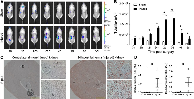
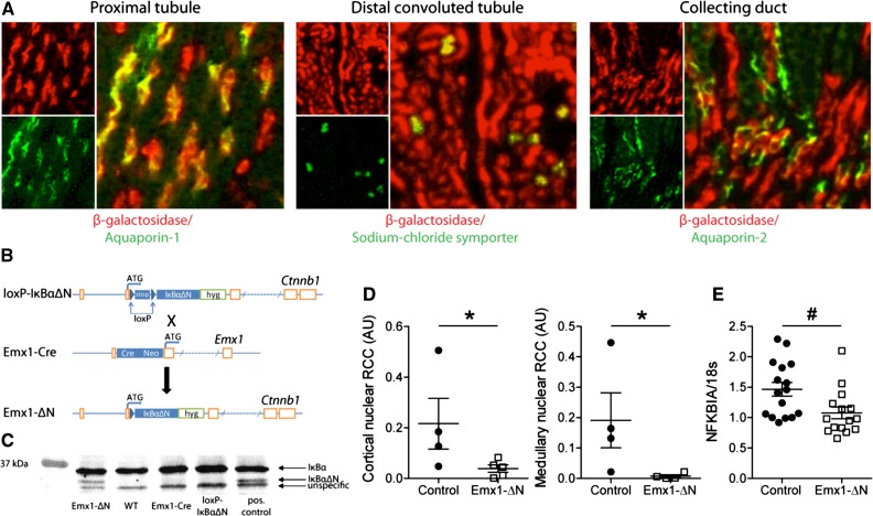
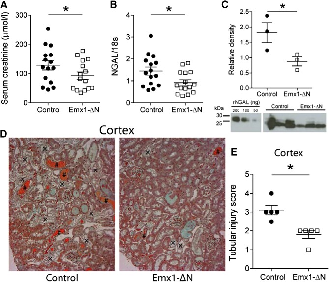
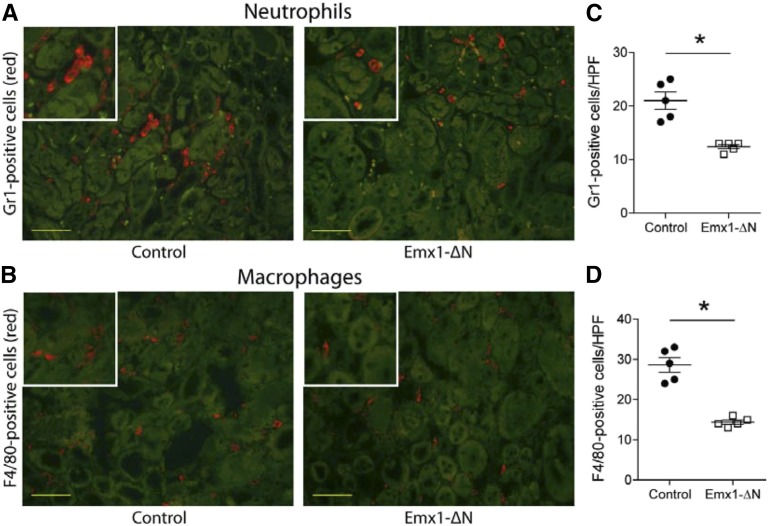
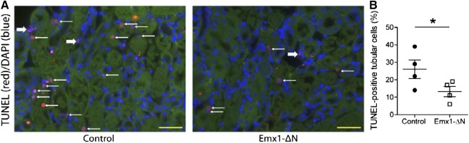
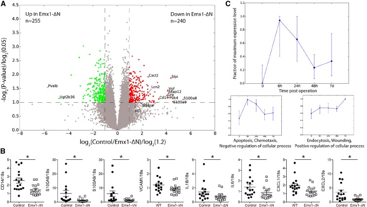
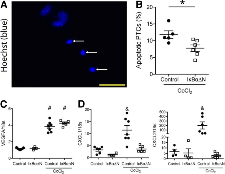
NF-κB is a key regulator of innate and adaptive immunity and is implicated in the pathogenesis of AKI. The cell type-specific functions of NF-κB in the kidney are unknown; however, the pathway serves distinct functions in immune and tissue parenchymal cells. We analyzed tubular epithelial-specific NF-κB signaling in a mouse model of ischemia-reperfusion injury (IRI)-induced AKI. NF-κB reporter activity and nuclear localization of phosphorylated NF-κB subunit p65 analyses in mice revealed that IRI induced widespread NF-κB activation in renal tubular epithelia and in interstitial cells that peaked 2-3 days after injury. To genetically antagonize tubular epithelial NF-κB activity, we generated mice expressing the human NF-κB super-repressor IκBαΔN in renal proximal, distal, and collecting duct epithelial cells. Compared with control mice, these mice exhibited improved renal function, reduced tubular apoptosis, and attenuated neutrophil and macrophage infiltration after IRI-induced AKI. Furthermore, tubular NF-κB-dependent gene expression profiles revealed temporally distinct functional gene clusters for apoptosis, chemotaxis, and morphogenesis. Primary proximal tubular cells isolated from IκBαΔN-expressing mice and exposed to hypoxia-mimetic agent cobalt chloride exhibited less apoptosis and expressed lower levels of chemokines than cells from control mice did. Our results indicate that postischemic NF-κB activation in renal tubular epithelia aggravates tubular injury and exacerbates a maladaptive inflammatory response.

Keywords: NF-kappaB; acute renal failure; apoptosis; chemokine; epithelial cells; ischemia-reperfusion; renal tubular.


Figures

Figure 1.
Figure 1.
Time course and localization of NF-κB activity in AKI. (A) In vivo bioluminescence whole-body imaging of κ-Luc mice following right uninephrectomy and sham surgery (control; upper panel) or 17.5 minutes of left renal ischemia (lower panel). After the procedure, mice were imaged at time points stated below. (B) NF-κB–driven luciferase activity in sham-operated mice (white bars; n=3) and in mice with ischemic kidney injury (black bars; n=4). nControl=15, nEmx1-ΔN=15; *P<0.05, Student's t test. p/s, photons/sec. (C) Immunostaining of contralateral kidney (noninjured; left panel) and of kidney 24 hours after ischemia (injured; right panel) with an antibody against phosphorylated (P) p65. Cd, collecting duct; Dt, distal tubule; G, glomeruli; Pt, proximal tubule. Black frames indicate sites of ×400 magnification. Yellow bars represent 1000 μm, and black bars represent 50 μm. (D) Quantification of nuclear P-p65 staining in cortex (left panel) and medulla (right panel). RCC, red channel count (see Concise Methods). nContralateral=4, nInjured=4; #P<0.05, Mann–Whitney U test.
Figure 2.
Figure 2.
Generation and characterization of a mouse model with renal tubule–specific expression of the human NF-κB super-repressor IκBαΔN. (A) Immunostaining on kidney sections from Emx1Cre;R26R mice using antibodies against β-galactosidase and aquaporin-1, sodium chloride symporter or aquaporin-2 to verify renal tubular Cre activity. (B) The cDNA of the NF-κB super-repressor IκBαΔN was knocked into the β-catenin (Ctnnb1) locus preceded by a loxP-stop-loxP cassette (loxP-IκBαΔN). Mating of loxP-IκBαΔN with Emx1-Cre knockin animals generated mice expressing IκBαΔN only in cells with an active Emx1 promoter (Emx1-ΔN). (C) Verification of renal IκBαΔN expression in Emx1-ΔN mice by Western blotting using an anti-IκBα antibody. Kidney tissue lysates isolated from IκBαΔNubi mice, which constitutively express IκBαΔN ubiquitously, served as positive control. (D) Quantification of nuclear P-p65 staining in cortex (left panel) and medulla (right panel) from control and Emx1-ΔN mice 24 hours after ischemia. RCC, red channel count (see Concise Methods). ncontrol=4, nEmx1-ΔN=5; *P<0.05, Mann–Whitney U test. (E) Expression of NFKBIA mRNA in control and Emx1-ΔN kidneys after 24-hour ischemia. ncontrol=16, nEmx1-ΔN=15; #P<0.05, t test.
Figure 3.
Figure 3.
Suppression of NF-κB activity in renal tubules ameliorates AKI. (A) Serum creatinine levels and (B) mRNA levels of kidney injury marker NGAL in Emx1-ΔN mice and control littermates 24 hours after ischemia. nControl=15, nEmx1-ΔN=15; *P<0.05, t test. (C) Urinary Western blot for analysis of NGAL expression using protein lysates from Emx1-ΔN mice and littermate controls. Recombinant mouse NGAL was used as a standard. nControl=3, nEmx1-ΔN=3; *P<0.05, t test. (D) Representative images of Masson trichrome stain on kidney sections of controls and Emx1-ΔN mice after 24 hours of ischemia (original magnification, ×400). Tubular necrosis (×) and eosinophilic cellular debris (#) are shown. (E) Semiquantification of cortical tubular injury. nControl=5, nEmx1-ΔN=5; *P<0.05, t test.
Figure 4.
Figure 4.
Inhibition of NF-κB activity in renal tubules is associated with reduced cellular infiltration after AKI. Immunofluorescence staining for neutrophil granulocyte marker Gr1 (red [A]) and for dendritic cell/macrophage marker F4/80 (red [B]) on representative kidney sections of control and Emx1-ΔN mice showing the outer medulla 24 hours after ischemia. Green staining is due to autofluorescence of tubules. Yellow bars represent 50 μm. Quantification of Gr1-positive (C) and F4/80-positive (D) cells. Each dot represents a single animal and is the mean of 20 randomly selected high-power fields (HPFs). nControl=5, nEmx1-ΔN=5; *P<0.05, t test.
Figure 5.
Figure 5.
Inhibition of NF-κB activity in renal tubules leads to reduced apoptosis upon AKI. (A) Representative ×400 images of TUNEL labeling on control and Emx1-ΔN kidney sections 24 hours after renal ischemia. Thin white arrows indicate TUNEL-positive tubular cells. Thick white arrows indicate TUNEL-positive interstitial cells. Yellow bars represent 50 μm. (B) TUNEL labeling was quantified by direct counting of the number of positively stained tubular cells, which was divided by the total number of cells per ×630 field. Each dot represents a single animal and is the mean of five randomly selected outer medulla fields per animal. nControl=4, nEmx1-ΔN=4; *P<0.05, t test.
Figure 6.
Figure 6.
Microarray analysis comparing the gene expression profile of control versus Emx1-ΔN kidneys after ischemia. (A) Volcano plot for genes obtained from microarray analysis. Axes show logarithmic transformation of fold changes (x-axis) and P values (y-axis). Genes differentially expressed (fold change ≥1.2 and P<0.05) are in green if upregulated in Emx1-ΔN mice compared with controls and in red if downregulated. (B) Verification by quantitative real-time PCR of some of the NF-κB target genes downregulated in injured Emx1-ΔN kidneys. nControl=15, nEmx1-ΔN=15; WT, wild-type. *P<0.05, t test. (C) Time-dependent expression pattern of all genes downregulated in injured kidneys of Emx1-ΔN mice compared with controls (red dots in A) at 0, 6, 24, and 48 hours and 7 days after ischemia. Displayed are time points 0, 6, 24, and 48 hours and 7 days. The upper panel shows median and quartiles for all differentially expressed genes downregulated in Emx1-ΔN compared with control, while the lower two panels are result of k-means clustering of these genes with respect to their timewise expression pattern (see Concise Methods for details). For each cluster, the dominant gene ontology (GO) term from gene ontology analysis and representative class members is depicted (for a full list of all members of each cluster and detailed result of GO analysis, see Supplemental Tables 1 and 2). All gene expression values have been maximum normalized for each gene.
Figure 7.
Figure 7.
Primary proximal tubular cells (PTCs) overexpressing IκBαΔN show less apoptosis and reduced chemotactic cytokine expression after CoCl2 treatment. (A) Representative image of apoptotic PTCs (white arrows; original magnification, ×630). Yellow bar represents 50 μm. (B) Quantification of apoptotic PTCs after 24-hour treatment of 300 μM hypoxia-mimetic agent CoCl2. Data are from two independent experiments. nControl=5, nIκBαΔN=6; *P<0.05, t test. mRNA expression of (C) HIF1α target gene VEGFA and (D) chemokines CXCL1 and CXCL2. Each dot represents a pool of PTCs isolated from two to three animals of the same genotype. Data are from two independent experiments. nControl=6, nIκBαΔN=4, nControl+CoCl2=7, and nIκBαΔN+CoCl2=6; #P<0.05 versus wild-type and IκBαΔN; and P<0.05 versus wild-type, IκBαΔN, and IκBαΔN+CoCl2. One-way ANOVA, Newman–Keuls multiple comparison post hoc test.

References

    1. Mehta RL, Cerdá J, Burdmann EA, Tonelli M, García-García G, Jha V, Susantitaphong P, Rocco M, Vanholder R, Sever MS, Cruz D, Jaber B, Lameire NH, Lombardi R, Lewington A, Feehally J, Finkelstein F, Levin N, Pannu N, Thomas B, Aronoff-Spencer E, Remuzzi G: International Society of Nephrology’s 0by25 initiative for acute kidney injury (zero preventable deaths by 2025): A human rights case for nephrology. Lancet 385: 2616–2643, 2015 - PubMed
    1. Bellomo R, Kellum JA, Ronco C: Acute kidney injury. Lancet 380: 756–766, 2012 - PubMed
    1. Glodowski SD, Wagener G: New insights into the mechanisms of acute kidney injury in the intensive care unit. J Clin Anesth 27: 175–180, 2015 - PubMed
    1. Ghosh S, Hayden MS: Celebrating 25 years of NF-κB research. Immunol Rev 246: 5–13, 2012 - PMC - PubMed
    1. Hinz M, Arslan SC, Scheidereit C: It takes two to tango: IκBs, the multifunctional partners of NF-κB. Immunol Rev 246: 59–76, 2012 - PubMed

Publication types